Eye-popping winning pictures from Nikon's Small World contest
From a bee's eye to carnivorous plants and a mouse colon, Nikon's photomicrography competition brings the small details of our world into startling focus.


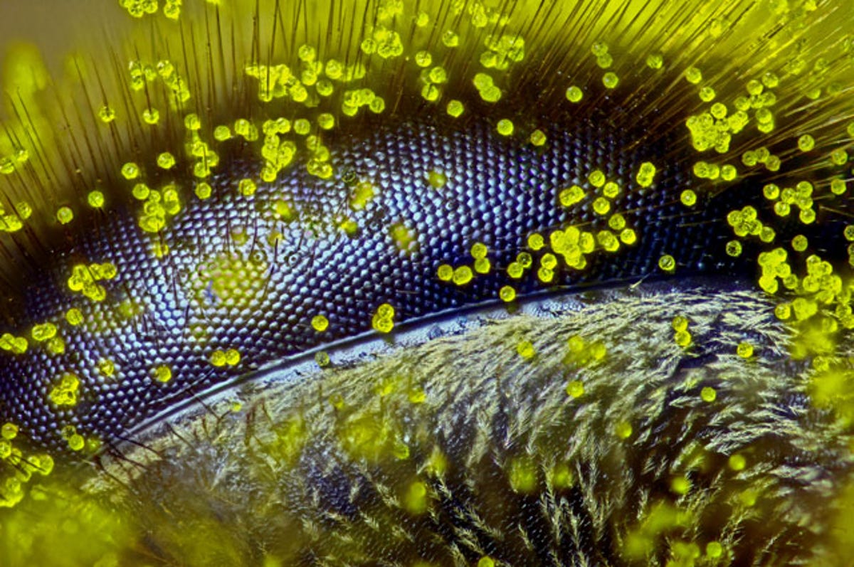
Well, eye bee!
Nikon this week announced the winners of its 41st annual Small World Photomicrography Competition recognizing images of the supersmall taken through microscopes.
"To select the winners, competition judges analyzed entries from all over the world covering subjects ranging from chemical compounds to up-close-and-personal looks at biological specimens," according to a statement.
More than 2,000 entries came in from over 83 countries. This shot of a bee's eye covered in dandelion pollen from Australian Ralph Grimm took first place. The image represents a zoom of 120x and dovetailed with Grimm's interest in beekeeping and the worldwide honey bee crisis.
"The aim of this image is to communicate to the viewer through the eye of a bee how intricate and beautiful nature is and that we are living in a super-busy high-tech world where an increasing number of people are losing their human identity in a steady decline of art as an important part of our society," Nikon said.
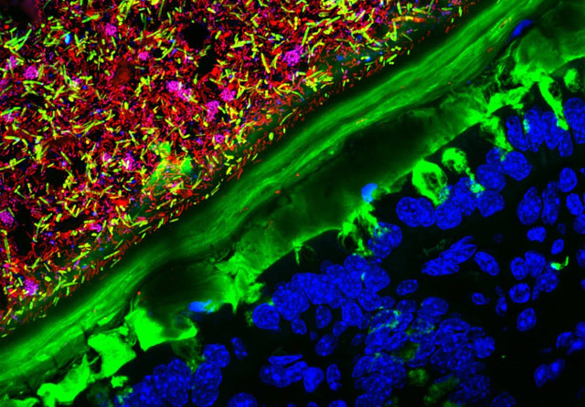
Colorful colony
It's hard to believe something as unattractive sounding as a mouse colon could look this vibrant, but that's exactly what this image shows. It won second place.
Specifically, it shows a mouse colon that started out completely germ-free and was then colonized with human bacteria. The image was shot using polarized light and 63x magnification.
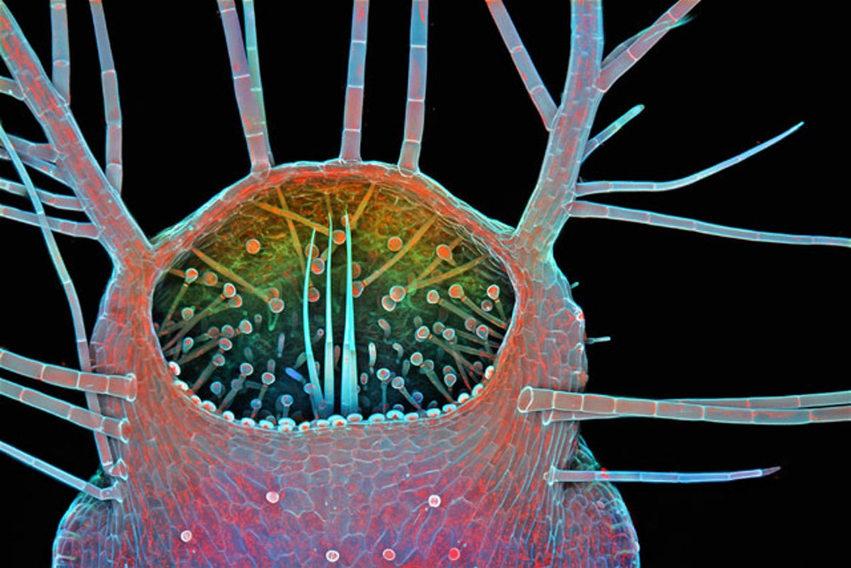
Humped bladderwort mouth
The humped bladderwort is a carnivorous plant that lives in freshwater. Here, 100x magnification shows the entrance to the protein-digesting plant.
"The bladderwort's trap is one of the most, if not the most, sophisticated plant organs in existence," Nikon said.
The photographer, Igor Siwanowicz, is a research specialist at the Janelia Research Campus of the Hughes Medical Institute. He's currently working on the steering circuit of a dragonfly.
"He thinks the relationship between science and art is best described in words of the French philosopher of science Jules Henri Poincare," Nikon said. "'The scientist does not study nature because it is useful; he studies it because he delights in it, and he delights in it because it is beautiful.'"
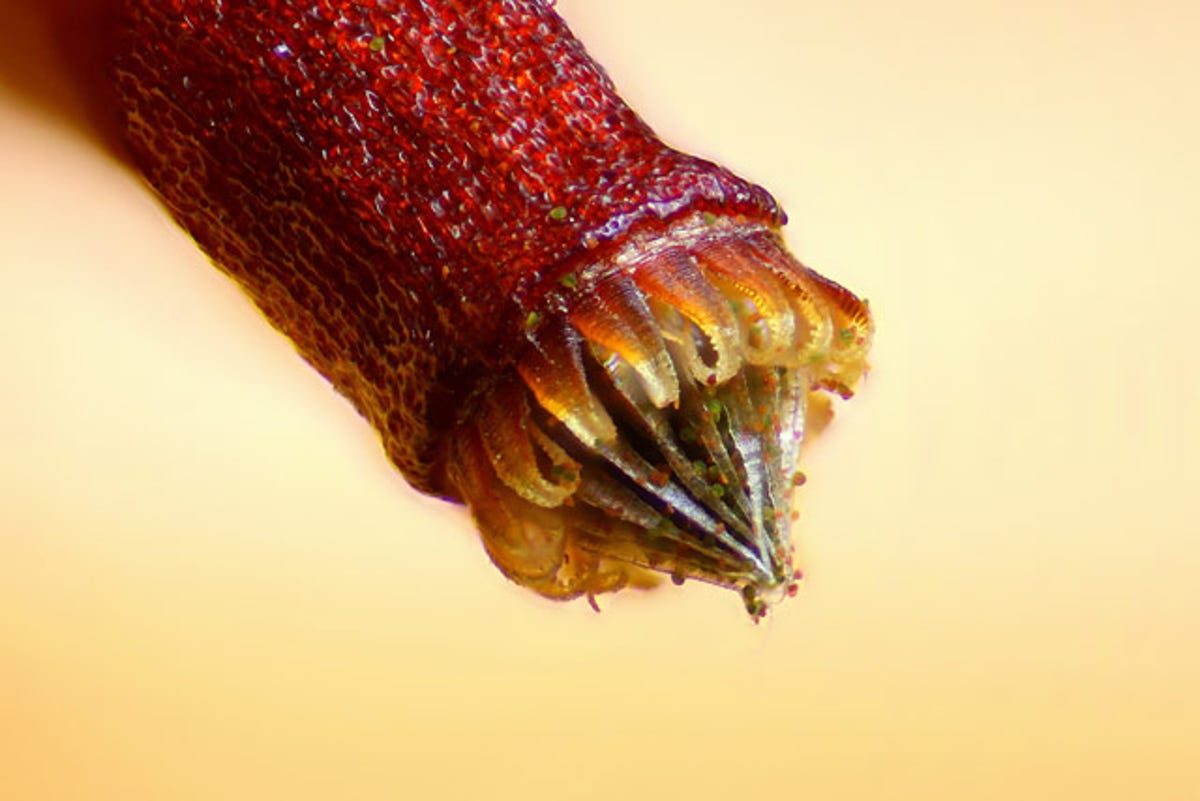
Sandworm?
In sci-fi writer Frank Herbert's "Dune" series, giant and fierce sandworms live beneath the surface of desert planet Arrakis. While this image certainly evokes those fearsome creatures, its origin is much more benign. It's simply a close-up shot of the spore capsule of a moss.
See? Harmless. But I bet you'll think twice before walking across a carpet of moss barefoot again, eh?
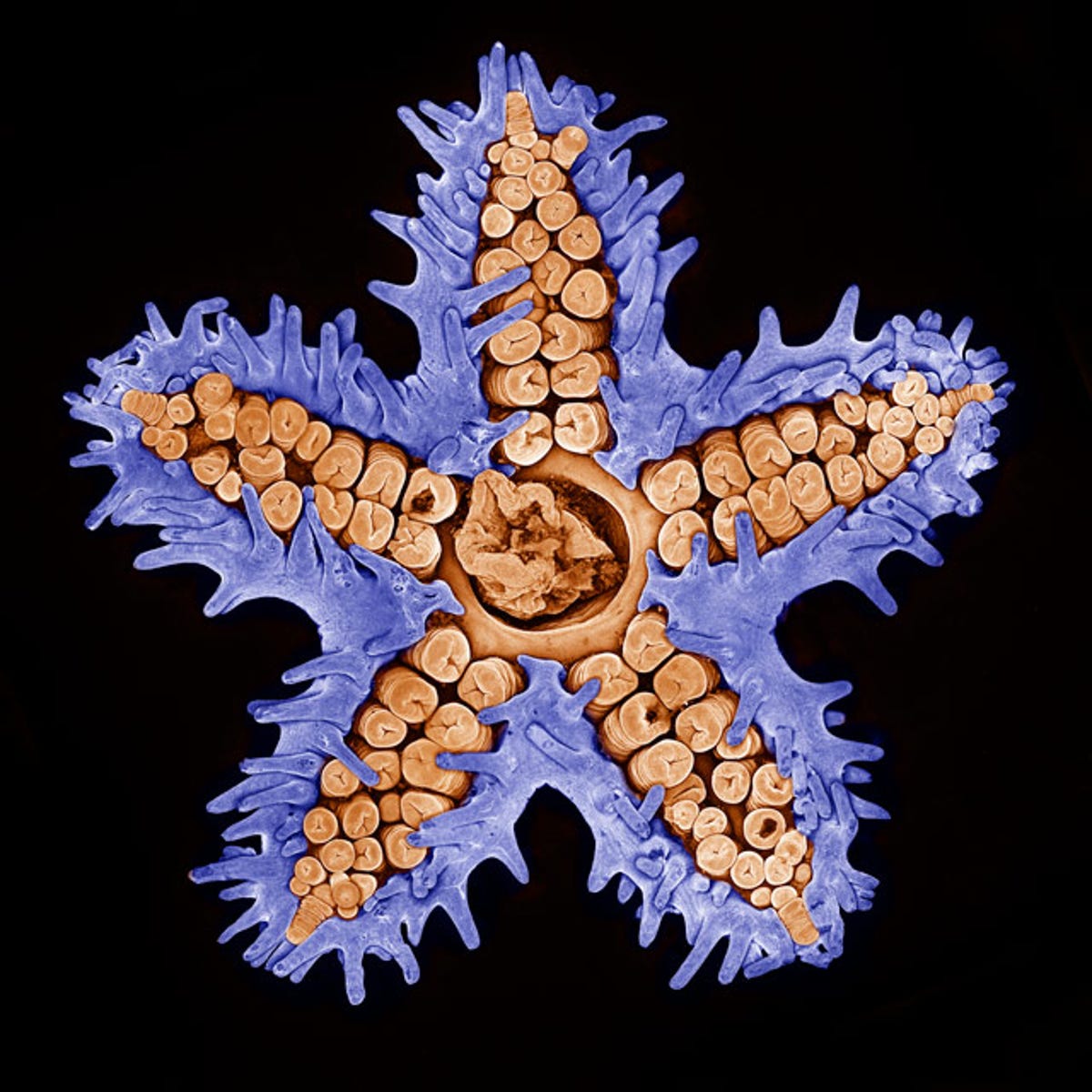
Superstarfish
In this image, an ordinary starfish looks spectacular under 10x magnification.
To create this shot, Evan Darling of the Memorial Sloan Kettering Cancer Center in New York used a technique called confocal microscopy. The technique involves scanning the specimen to create slices that are as thin as 250 nanometers (about 100 times thicker than a human hair). The slices are then stacked to produce a 3D image of the specimen.
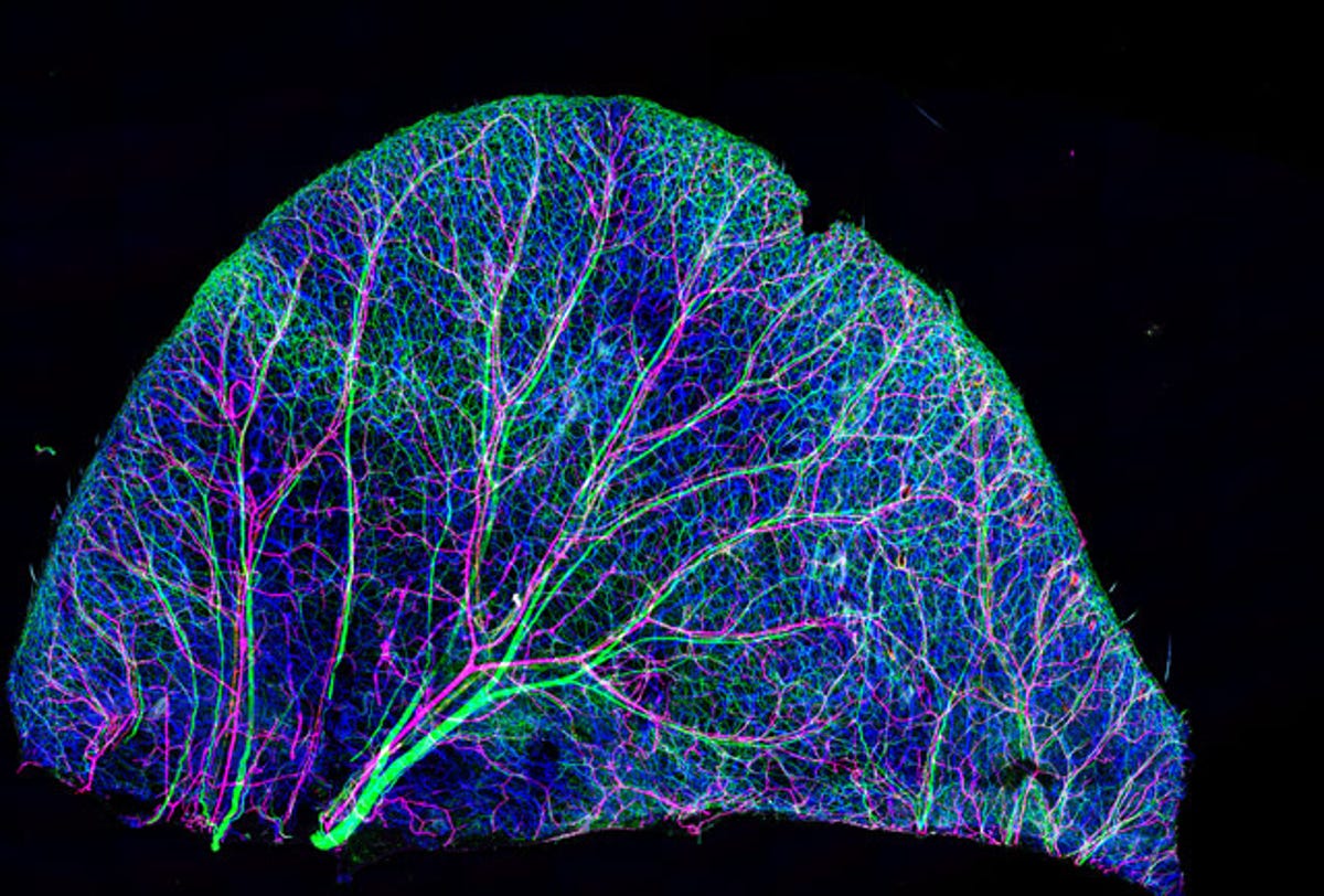
Animal, vegetable or mineral?
On first glance, this image looks like a leaf. It is, however, a look at the nerves and blood vessels in mouse ear skin under a 10x magnification.
Tomoko Yamazaki of the National Institutes of Health created the photo using confocal microscopy.

Best buds
The confocal technique came into play again for this ninth-place entry taken by Nathanaël Prunet from Caltech. The shot shows the young buds of a flowering plant called arabidopsis under 40x magnification.
Arabidopsis is often used in studying biology and has been entered many times in the Nikon contest. It is the first plant to have had its entire genome sequenced.
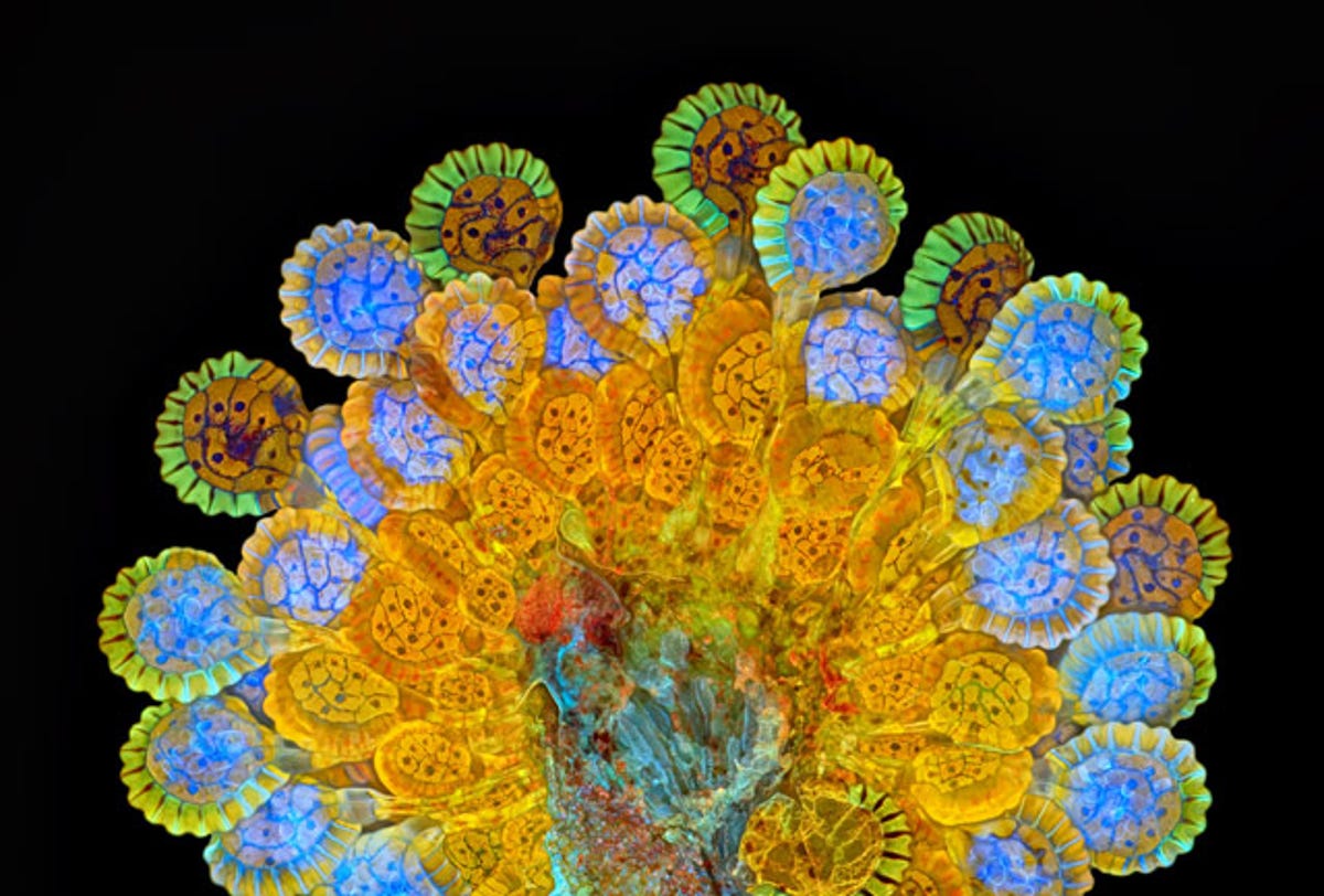
Fantastical fern
Though it looks like a lollipop from Willy Wonka's candy factory, this image actually shows a fern's sorus at different stages of maturity. A sorus is a collection of spore-producing structures usually found on the underside of fern fronds.
To create the image, photographer Rogelio Moreno Gill from Panama used fluorescence and image stacking. "Fluorescence imaging uses high-intensity illumination to excite fluorescent molecules in the sample," Nikon explained.
Image stacking is a technique wherein multiple shots are taken of a subject at different f-stops and the sharpest parts from each shot are compiled into one image.
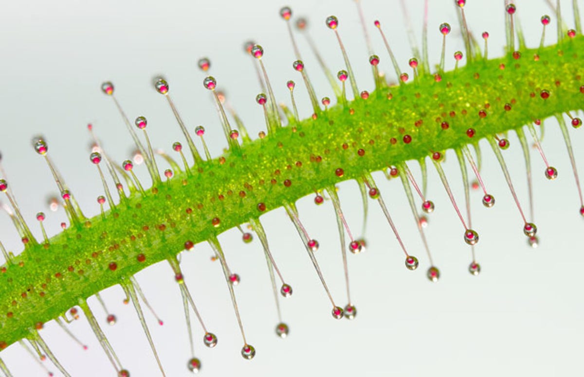
Good enough to eat?
This shot of a plant part looks good enough to eat. But if you were a small insect, chances are better it would eat you.
The shot of a tentacle from the carnivorous plant drosera, or sundew, was created using image stacking by José R. Almodóvar of the University of Puerto Rico.
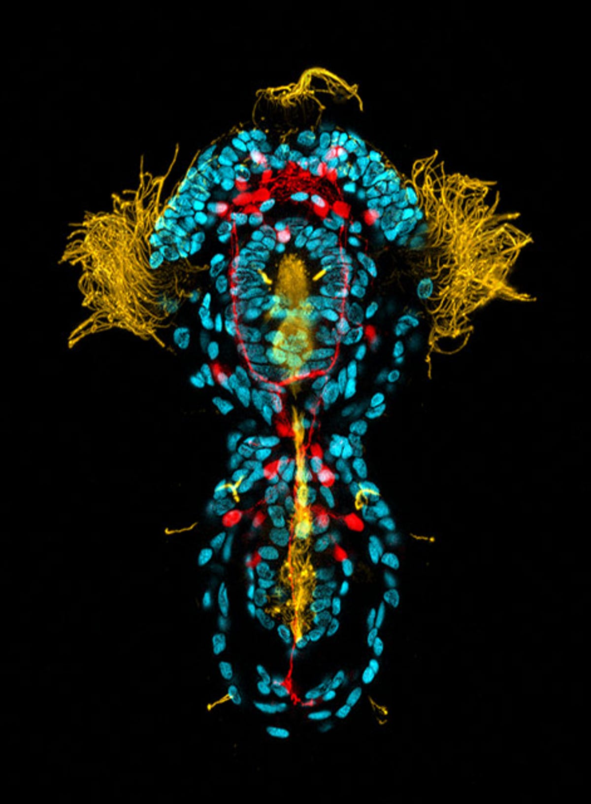
Wondrous worm
This image, which won an honorable mention in Nikon's contest, shows the 3-day-old larva of a peanut worm under 40x magnification.
The yellow represents the creature's cilia, the blue its DNA and the red shows the serotonin in its nervous system. The image was created by Michael J. Boyle of the Smithsonian Marine Station.
More Galleries

My Favorite Shots From the Galaxy S24 Ultra's Camera
20 Photos
Honor's Magic V2 Foldable Is Lighter Than Samsung's Galaxy S24 Ultra
10 Photos
The Samsung Galaxy S24 and S24 Plus Looks Sweet in Aluminum
23 Photos
Samsung's Galaxy S24 Ultra Now Has a Titanium Design
23 Photos
I Took 600+ Photos With the iPhone 15 Pro and Pro Max. Look at My Favorites
34 Photos