Best medical science images of 2015
The Wellcome Trust has announced the 20 winners of its 2015 Image Awards for medical science images. Here are our favourites.


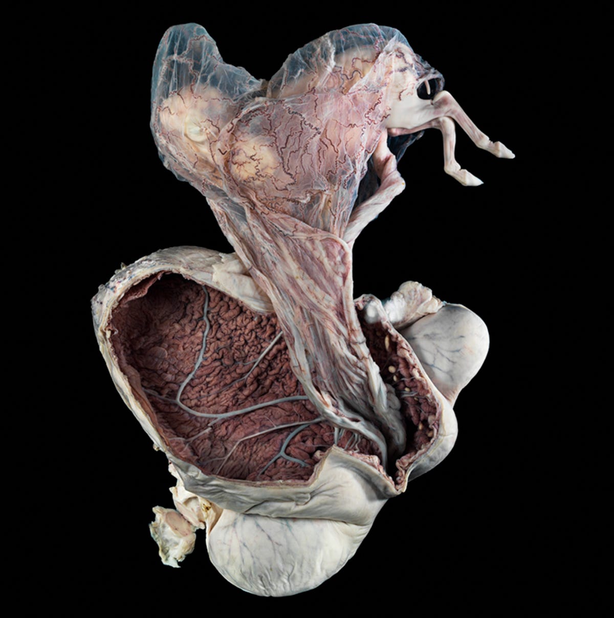
Grand prize winner: Pregnant uterus, equine
Snapped by photographer Michael Frank in collaboration with Nick Short of the Royal Veterinary College in London, this image shows the uterus of a New Forest pony preserved in formalin and kept in a Perspex container, complete with foetus at about five months into its 11-month gestational period, currently housed at the college's Lanyon Anatomy Museum. The membranes and umbilical cord still attach the foetus to the womb, which has been cut open to display the rich blood supply on the interior.
"As far as standout images go, the image of the horse's uterus with the foetus still inside was incredible and just sticks in my mind. It evokes many different emotions at once. It's fascinating, sad, macabre, almost brutal. Yet the subject is also delicate, detailed and beautiful," said Wellcome Image Awards judge James Cutmore, picture editor of BBC Focus magazine. "The image shows us a large and magnificent creature reduced to this sad, fragile and half-formed creation, which I find very humbling."
Click through the rest of the gallery for our favourite Wellcome 2015 Image Award winners, and visit the website for the full set.
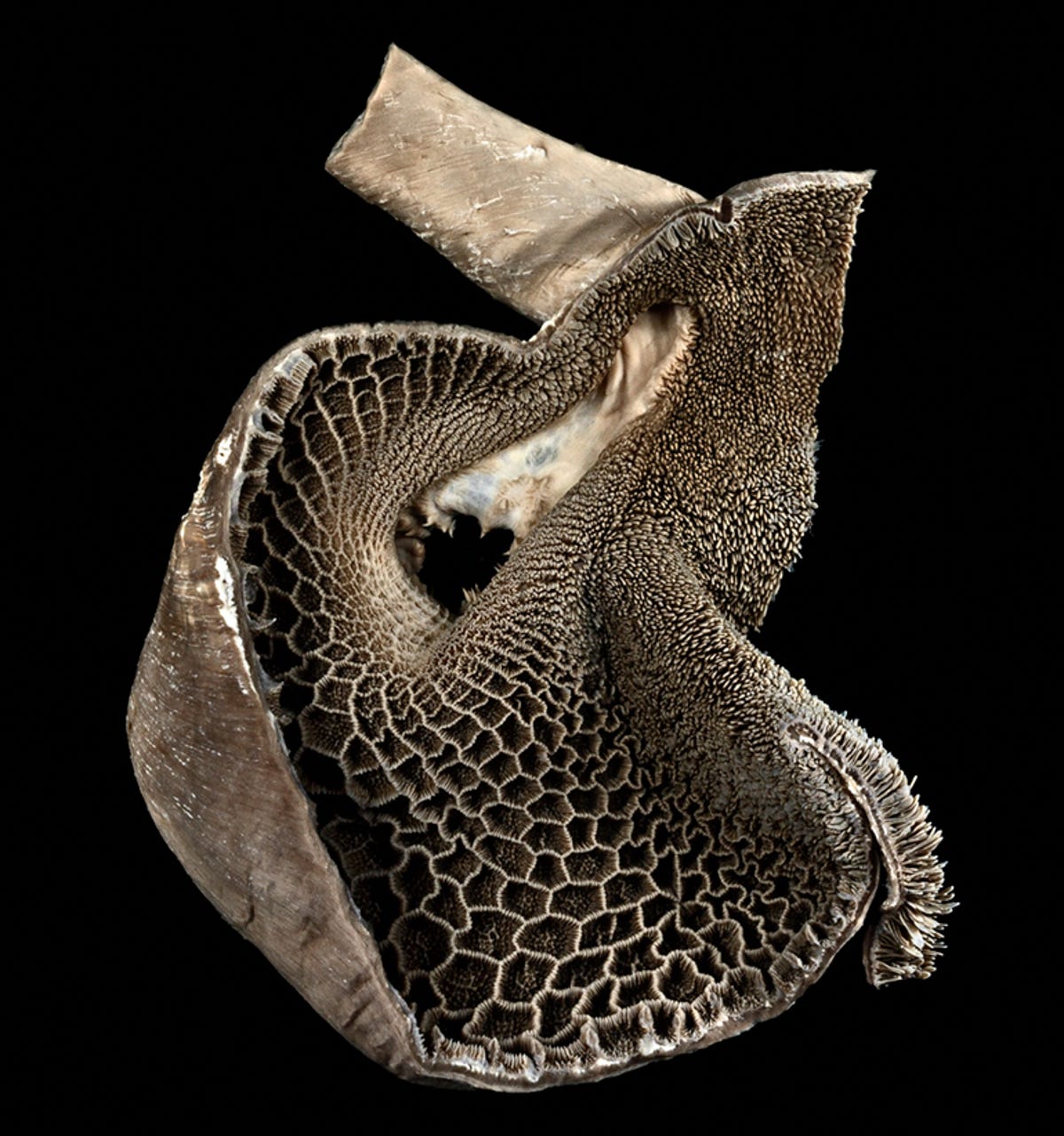
Goat stomach chamber
Michael Frank's collaboration with London's Royal Veterinary College saw several formalin-preserved specimens immortalised as stunning images. This image shows the reticulum -- the first of four chambers in the alimentary canal that make up the stomach of a ruminant animal -- in this case, a goat.
This chamber is also known colloquially as the "honeycomb" due to the shape of the internal mucosa; this part of the reticulum may be separated and eaten as tripe, but in its actual function, it is home to the stomach bacteria that help break down the animal's food.
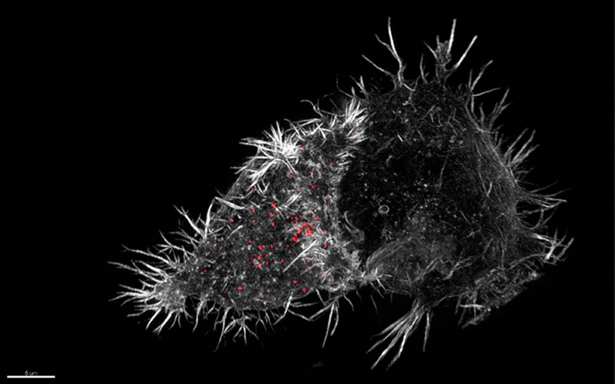
Active immune defense
A tale of two cells. On the right is a normal cell. On the left, latched to it, is what is known as a natural killer cell. These are the soldiers of the immune system, seeking out infected or cancerous cells and attacking them in order to keep the body healthy. This image, captured by biochemist Nele Dieckmann and microscopy specialist Nicola Lawrence, has caught this process in action.
The NK cell has attached itself to the normal cell and is scanning it for signs of disease, changes in the cell caused by infection, stress or malignancy -- and preparing to release chemicals -- the bright red dots -- that will cause the normal cell to self-destruct if it does turn out to be diseased.
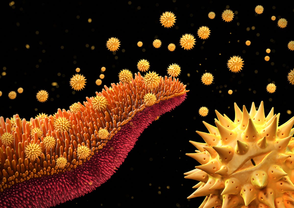
Pollen spores
Prolific science illustrator Maurizio De Angelis is behind this compelling illustration of the release of Asteraceae pollen spores -- the family of flowering plants to which daisies, asters and sunflowers belong. The microscopic spores are usually released in the spring and are a common allergen. However, they're also an integral part of the way plants reproduce.
The pollen on a flower's stamen contains the male sperm cells of the plant, which need to be transferred to the female reproductive structure -- the pistil -- in order to create a seed. This is primarily achieved through transport via insects such as bees and butterflies, birds and the wind.
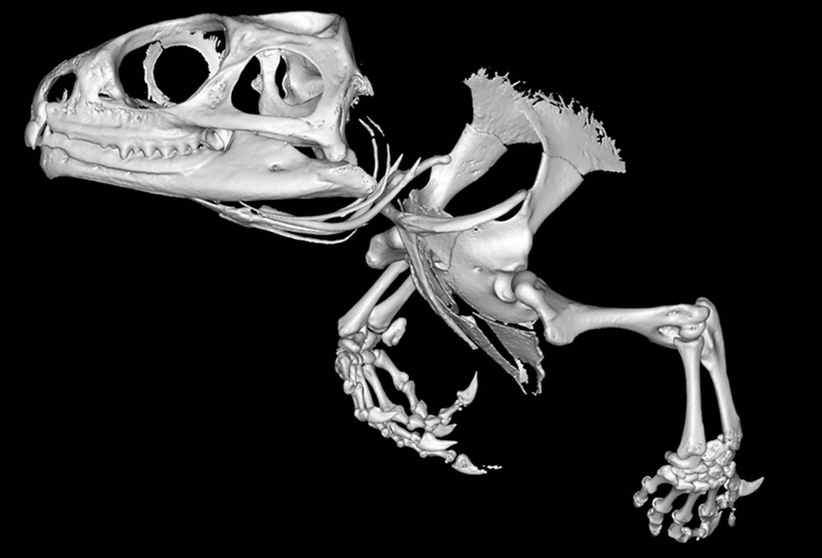
Tuatara
They look just like lizards, but the tuatara of New Zealand are something very special indeed -- the last surviving members of a family of reptiles that used to live with the dinosaurs. Although they live only in New Zealand now, 245 million years ago, tuatara populated the world.
The two remaining tuatara species -- named for their spiny crest, "tuatara" is a Maori word meaning "spiny back" -- have changed quite a bit from their prehistoric ancestors, which makes them a valuable study in evolution. This micro-computed tomography image shows the skull and front limbs of the tuatara, allowing a close study of the tiny bones within its tendons, usually hard to find in dissection; examining these and how they have changed will allow researchers to understand how these bones affect the tuatara's movement.
The art of a fruit fly
This is not a painting; in fact, if we were looking at this image at 1:1 scale, it would measure just 15 micrometers (0.015 millimetres) across. It's a digital colour-coded map of the nervous system of a fruit fly. The neurons that can sense vibrations are coloured yellow, while the blue and red circles represent information entering and leaving the synapses respectively. The orange circles represent mitochondria.
The image was created by taking electron miscroscopy images of very thin slices of the fly's tissue, which were then reconstructed to make a 3D model.
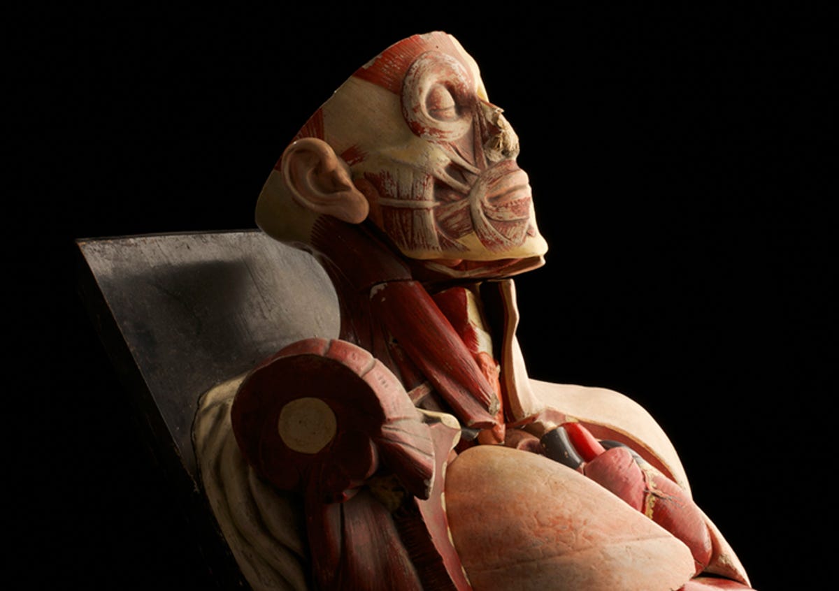
Tha anatomy model
After decades of service at the Trinity College, Dublin, this anatomical model on its way to the skip got a last hurrah from artist and photographer Anthony Edwards.
Awards judge and science broadcaster Adam Rutherford explained, "The model, the photo and the story are all beautiful. These models are the next best thing to actual bodies for learning anatomy, and have a kind of beauty about them. The fact that it was retrieved from a dump at Trinity makes its story tragic and we felt, like the photographer, that its service and beauty were worth honouring."
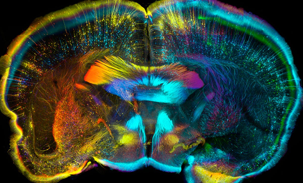
In the mind of a mouse
Confocal micography on a 0.75-millimetre thick slice of mouse brain here reveals the structures inside. The colour codes, created with chemicals to make the structures more visible, paint the closest nerve cells as reds and oranges, with the farthest as blues and greens. This technique is being used to map the wiring of entire brains.
"The beautiful colours and incredible level of detail in this image drew us in, inviting us to look more and more closely at it. The fine lines showing the nerve fibres 'shooting' across this tiny slice of brain also give it a feeling of movement and complex activity," awards judge and head of Wellcome Images Catherine Dracott said.
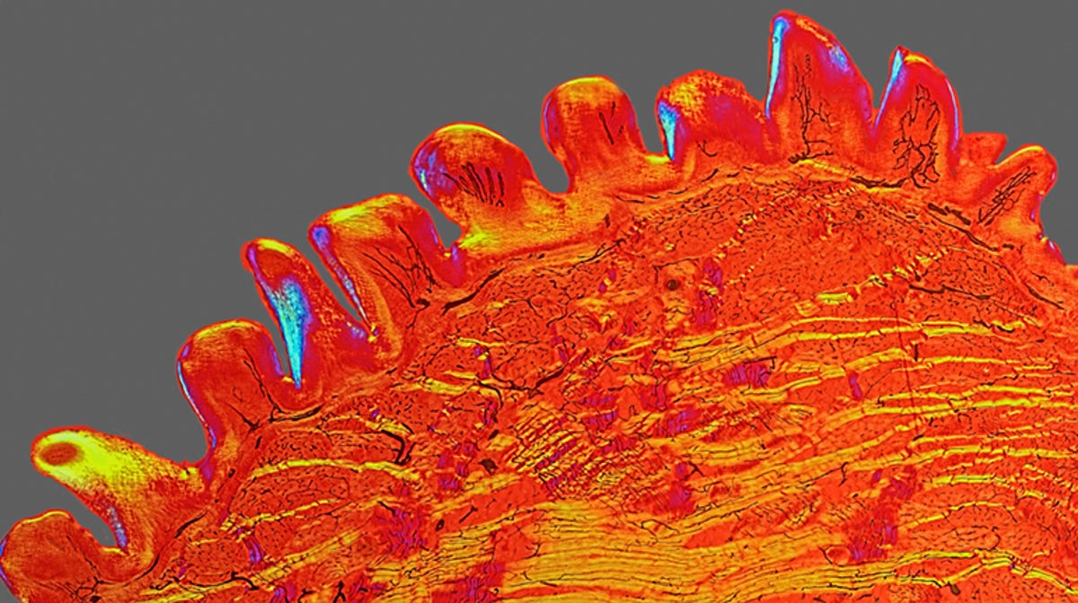
Rough as a cat's kiss
Cat owners know that sandpapery feeling when a cat decides you need a bit of a wash. This polarised light micrograph of a three-millimetre wide cross-section of cat's tongue shows the rough structure that creates that sandpapery feeling; although not as pleasant on skin, for a cat's fur, the structures act like a sort of comb, cleaning the fur and "brushing" it neatly into place.
This picture was taken from a very old slide, prepared in the Victorian era, using a technique that is well known now, but that was very new at the time. Black dyes were injected into the capillaries to make them more visible.
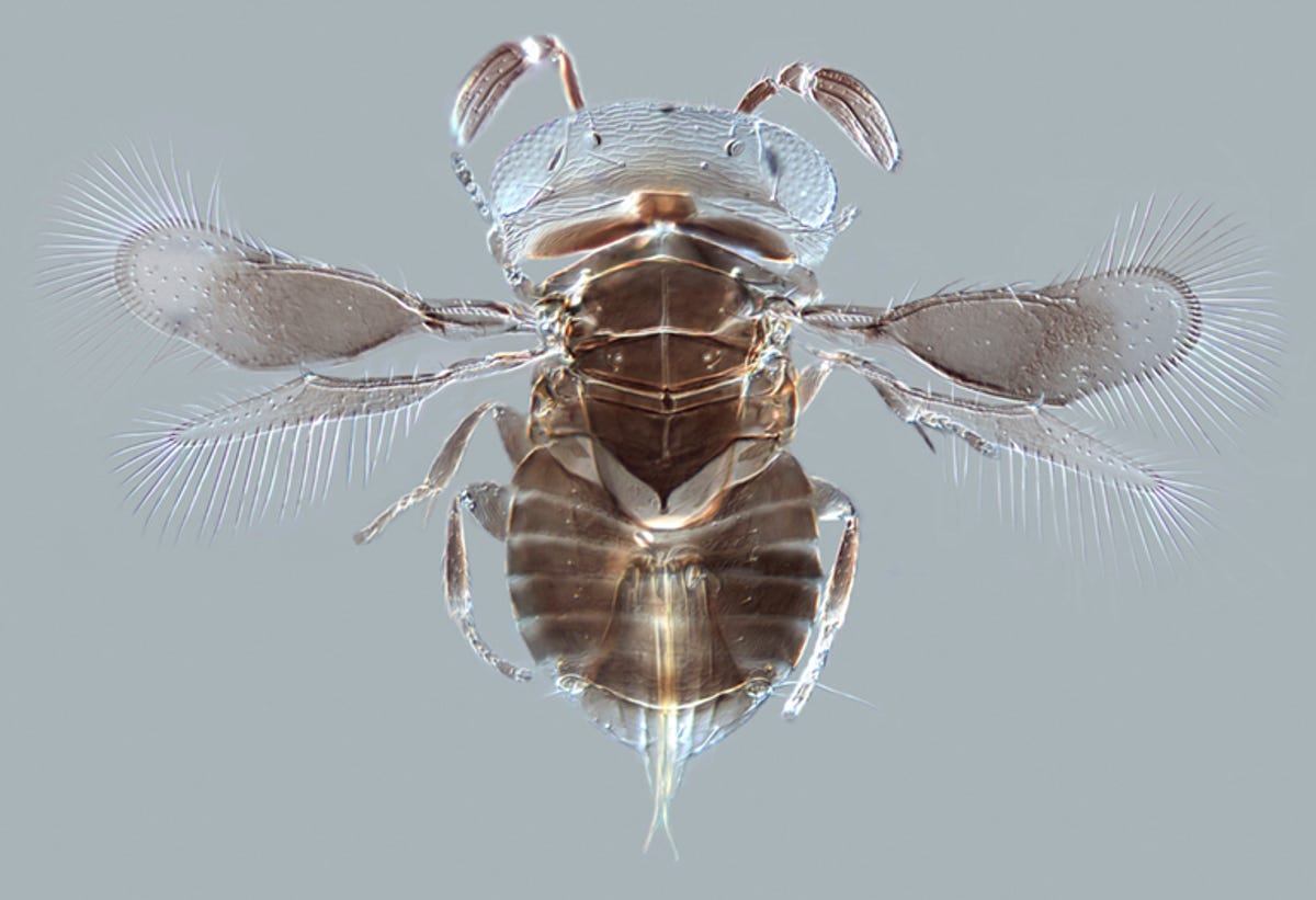
Parasitoid wasp
Parasitoid wasps are some of the most skin-crawly creatures in the insect kingdom. These wasps lay their eggs inside other insects; when the eggs hatch, they eat their way out of the still-living host from the inside. Blurgh. Although it may give you the willies, parasitoid wasps often prey on agricultural pests -- making them a highly effective natural pesticide.
This light micrograph shows a species recently discovered in the rainforests of Borneo -- a single female specimen mixed in with thousands of other insects. It measures just 0.75 millimetres in length; this photograph shows in detail the wasp's unusual legs, wings and antennae.
Visit the Wellcome Trust website for the rest of the award winners, and let us know your favourite on Twitter or Facebook.
More Galleries

My Favorite Shots From the Galaxy S24 Ultra's Camera
20 Photos
Honor's Magic V2 Foldable Is Lighter Than Samsung's Galaxy S24 Ultra
10 Photos
The Samsung Galaxy S24 and S24 Plus Looks Sweet in Aluminum
23 Photos
Samsung's Galaxy S24 Ultra Now Has a Titanium Design
23 Photos
I Took 600+ Photos With the iPhone 15 Pro and Pro Max. Look at My Favorites
34 Photos