Breakthrough in tissue engineering: 'Bio-Legos'
A new technique called micromasonry uses a gel-like material to bind cell bricks together as the material hardens, enabling the formation of 3D shapes to build such things as new organs.


Researchers have been working on the problem of tissue engineering for years because the payoff would be so great. The ability to construct new organs would mean that patients won't necessarily have to wait for transplants.
But growing cells in lab dishes that are three-dimensional instead of flat has proved to be incredibly tricky. One group, however, claims to have made a major breakthrough.
The solution, according to a team at the MIT-Harvard Division of Health Sciences and Technology (HST), could be as simple (but incredibly elegant) as building the equivalent of biological Legos, whose structures far more closely resemble real, complex tissue.
The new technique, which they're calling "micromasonry," is showcased this May in the journal Advanced Materials.
According to former HST postdoctoral associate Javier Gomez-Fernandez, obtaining single cells to engineer tissue requires breaking tissue apart via enzymes that digest the material that holds cells together. Once those cells are free they don't easily (or quickly) reassemble into structures that mimic natural tissue architecture.
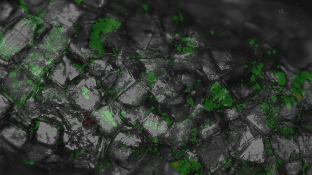
So the HST team built what it's dubbing "biological Legos," coating the freed cells with a liquid version of polyethylene glycol (PEG), which acts as a glue and also hardens when illuminated. Once coated, they could be arranged into cubes and exposed to light to hold that shape.
The team then coated those cubes with the PEG polymer again to glue the cubes together and squeeze them onto a scaffold surface, and illuminated the cubes a second time so that they would harden into this tube-like shape, a three-dimensional structure that could function as capillaries and transport blood to organs.
The breakthrough is not only that they are creating 3D structures, but that they can place cells in any position they want to make specific shapes. "So it's not just that it's a scaffold, it's that you can arrange them into very specific shapes through specific distributions of cells," Gomez-Fernandez says.
This isn't the first time complex tissue architecture has been created in a lab; other researchers have developed a technique called organ printing to accomplish this, but it requires new (read: expensive) equipment. Micromasonry, on the other hand, does not. "You can reproduce this in any lab," Gomez-Fernandez says in the MIT news release. "It's very simple."
To make micromasonry clinically useful, the team is investigating different cellular structures as well as different polymers that might provide more control over cell placement than PEG currently allows.