Color X-ray scanner uses CERN tech to see all your innards
Fat, bone, metal. It's all there in 3D.


The familiar black-and-white X-ray could soon be replaced with detailed 3D color scans that show everything from fat and bone to metal and soft tissue.
Phil and Anthony Butler, a father-and-son scientist team in New Zealand, created the MARS spectral x-ray scanner by adapting technology used at the European Organization for Nuclear Research (CERN). CERN is famous for hunting the Higgs boson, also known as the "God particle," with the Large Hadron Collider particle accelerator.
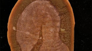
The 3D color scans deliver significantly more medical information than a conventional X-ray, showing fat, water, calcium and disease markers. The scans look like detailed 3D anatomical cross sections with the skin peeled away.
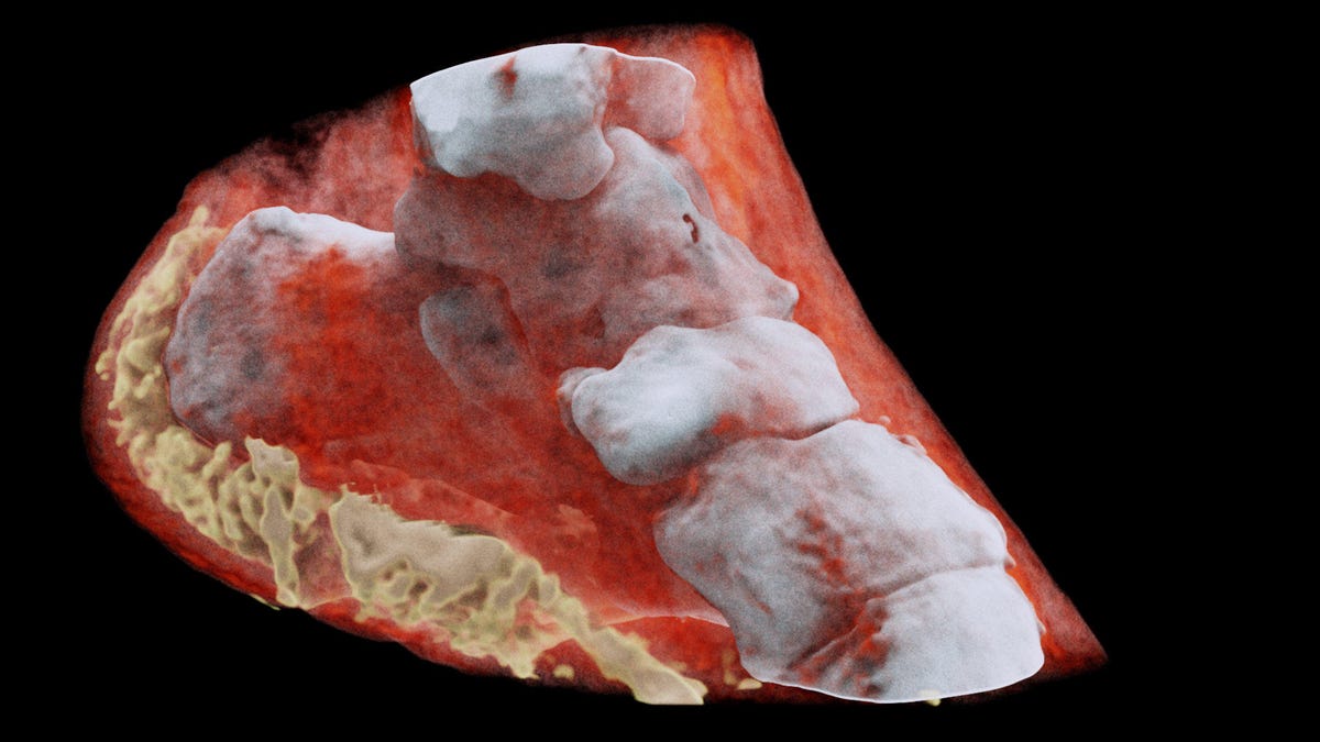
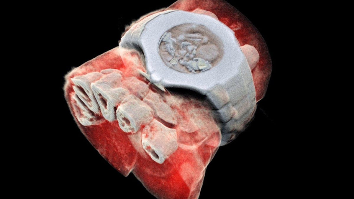
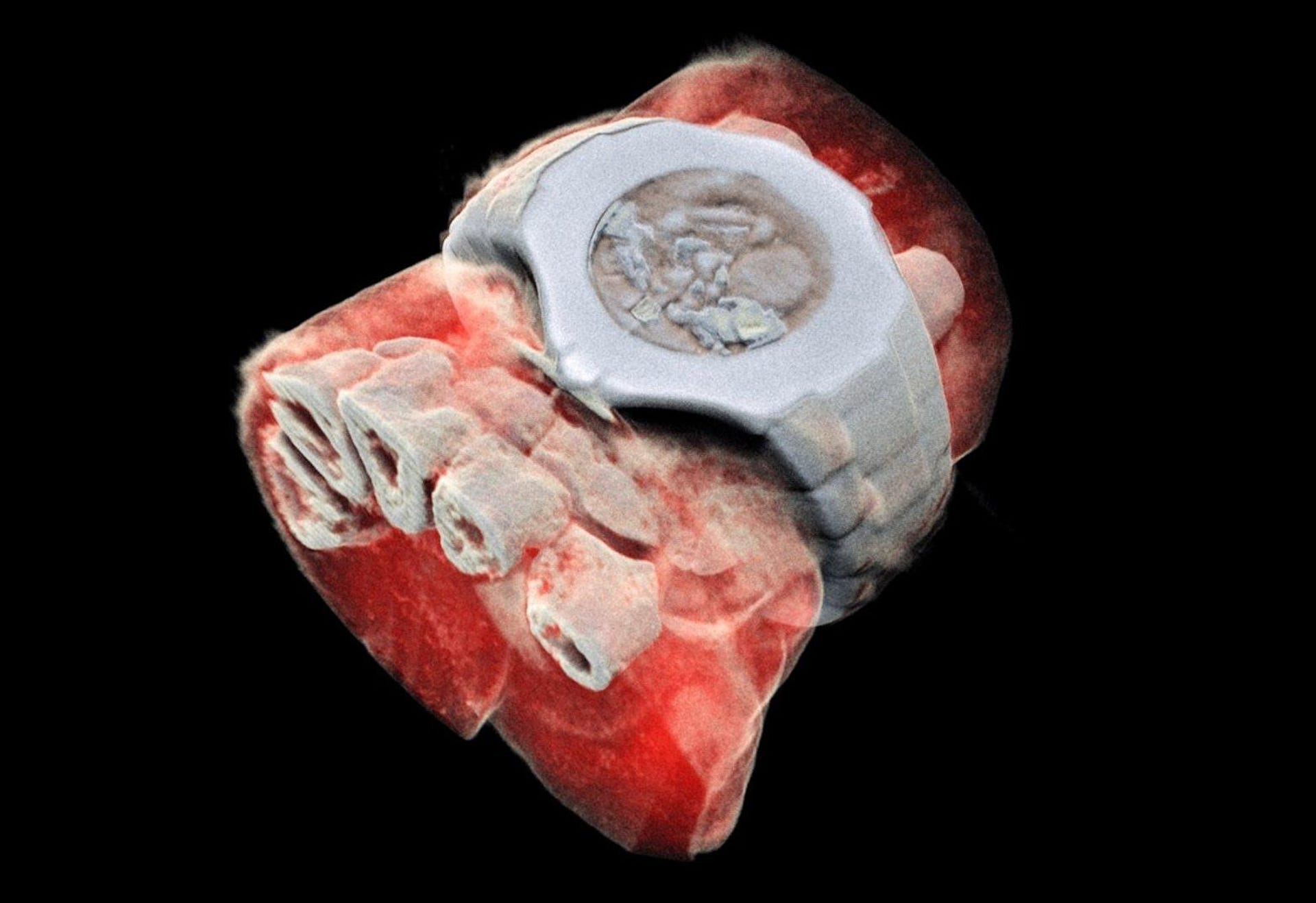
The MARS scanner produced this view of Phil Butler's ankle.
"So far researchers have been using a small version of the MARS scanner to study cancer, bone and joint health, and vascular diseases that cause heart attacks and strokes," Anthony Butler, a radiologist at the Universities of Otago and Canterbury, said in a press release.
The MARS scanner incorporates Medipix chip technology developed at CERN. "The original concept of Medipix is that it works like a camera, detecting and counting each individual particle hitting the pixels when its electronic shutter is open," CERN said in a statement. The technology results in "high-resolution, high-contrast, very reliable images," making it ideal for use in medical imaging.
MARS Bioimaging is commercializing the scanner. Phil Butler became the first person to be scanned with MARS, producing eye-opening images of his ankle and wrist. MARS Bioimaging is now moving on to clinical trials with scans of orthopedic and rheumatology patients.
Anthony Butler says the MARS scanner will enable more accurate diagnosis of medical conditions ranging from cancer to arthritis and will allow for more personalized treatment plans.