Skeletons shine under eerie new imaging techniques
It's "like a Grateful Dead poster," says one scientist.


A lot of scientific papers on animals include specimen photos that are very clinical in nature. You wouldn't mistake them for works of art. Until now.
New imaging methods developed by researchers at the University of Kansas in Lawrence are turning vertebrate innards into views that will dazzle your eyes.
The first method involves combining glycerine and gelatin into a see-through mixture that helps cleaned skeletons hold their shape for photographing.
"The problem we had was we couldn't pose these animals because we've digested away all of the muscles," said W. Leo Smith, associate professor of ecology and evolutionary biology. "They're flaccid and useless, like a pile of clothes that fold in every direction."

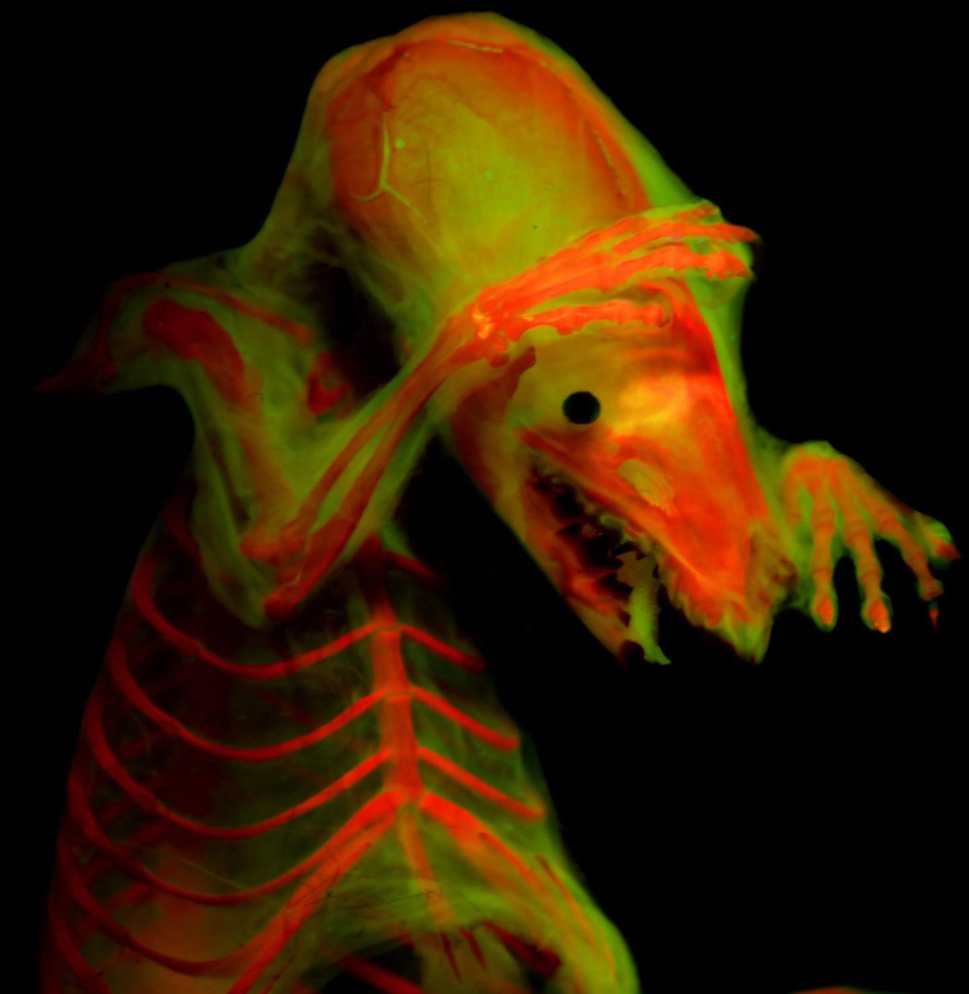
Cleared-and-stained North American Least Shrew under fluorescent light.
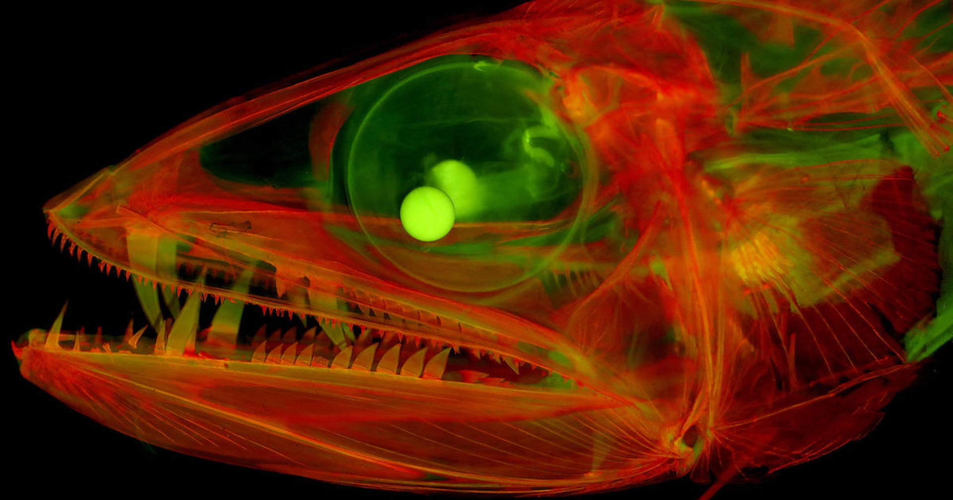
The glycerin-gelatin mix lets scientists photograph skeletons from various angles, enabling hard-to-capture views, such as a head-on look at a fish's teeth and jaws.
"There's something different about being able to see anatomical structure in new ways that really does help analysis," said Smith.
The second technique involves fluorescent microscopy and a stain called alizarin, which fluoresces under certain wavelengths of light.
"Alizarin red is used to dye a specimen's bones, and it fluoresces like a Grateful Dead poster," Smith said.
The method uses a microscrope equipped with filters that block out other light, leaving a bright image of fluorescing bones for the camera.
The research team's images of stained specimens treated with alizarin are truly stunning, showing the minute details of joints, tiny bones and teeth.
Smith hopes these techniques will help scientists better illustrate and communicate their research through more compelling images. The team published details on the new methods in the September issue of the journal Copeia.

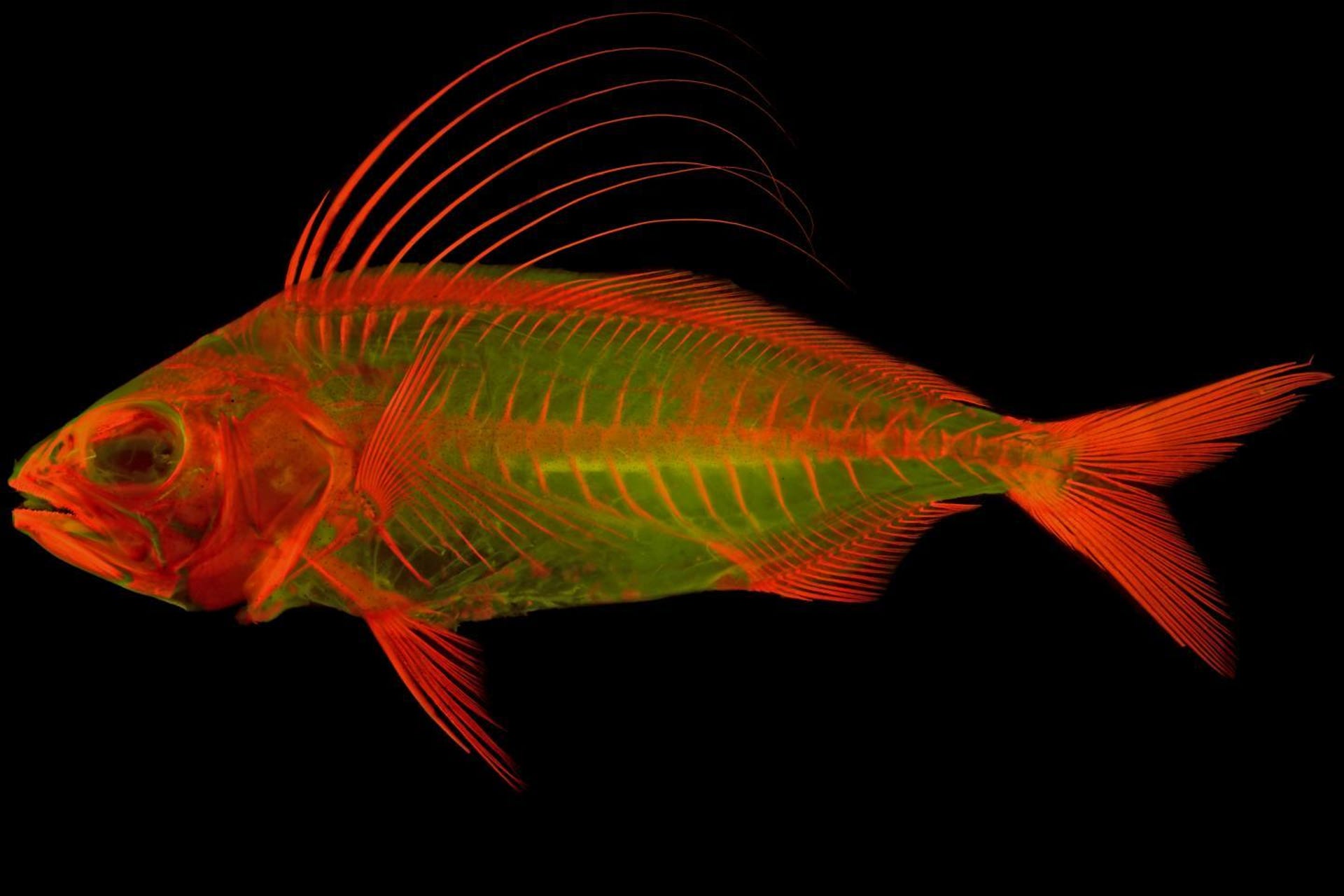
Stained juvenile Roosterfish under fluorescent light.