Miniature brain-in-a-dish could help advance Alzheimer's research
A lab-grown brain is the most complete ever developed, equivalent to the brain maturity of a five-week-old foetus.


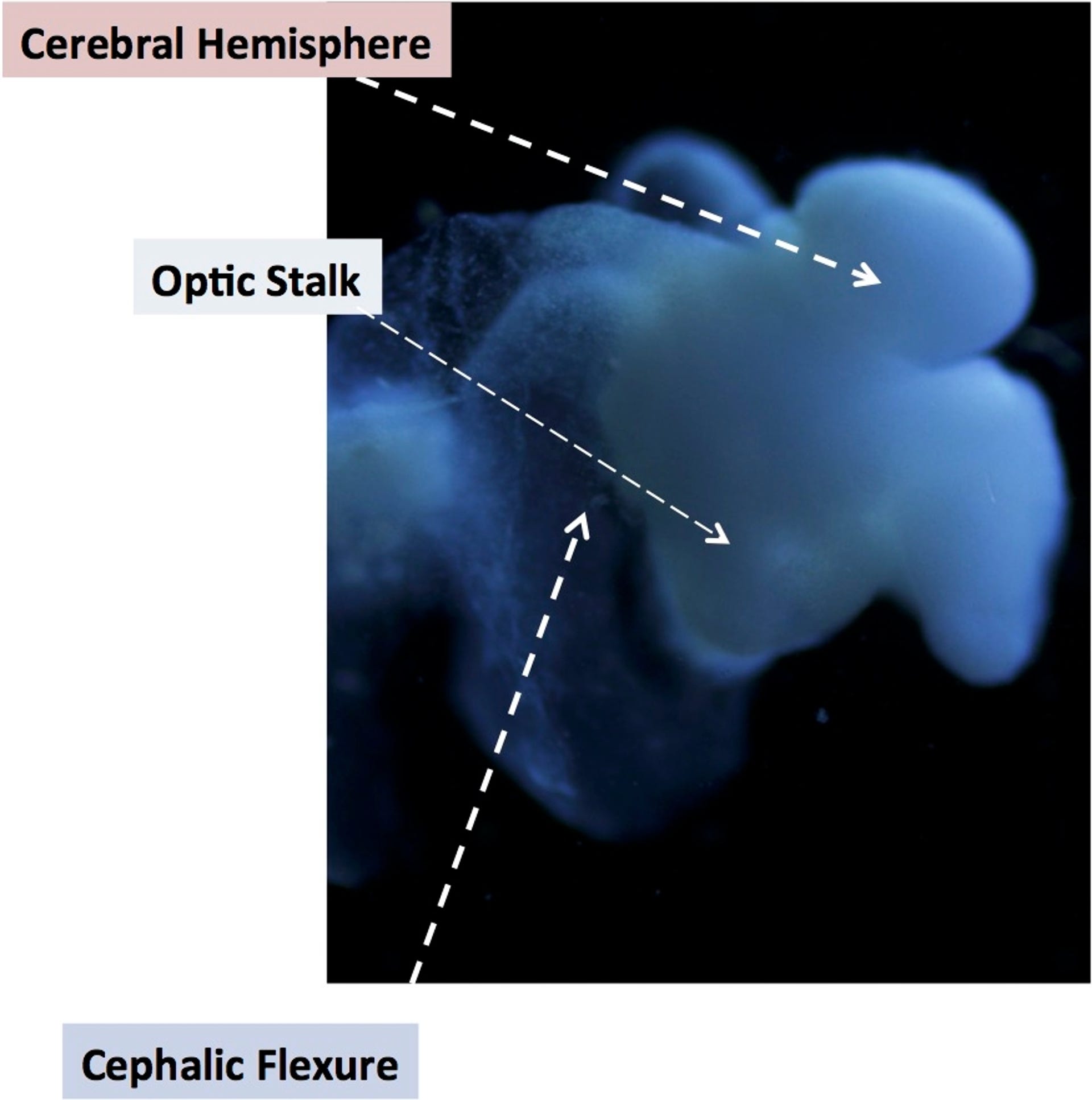
It's only about the size of a pencil eraser, but a lab-grown brain has big implications for the study of neurological disorders.
"It not only looks like the developing brain, its diverse cell types express nearly all genes like a brain," said Rene Anand, professor of biological chemistry and pharmacology at Ohio State University, who developed the organoid.
"We've struggled for a long time trying to solve complex brain disease problems that cause tremendous pain and suffering. The power of this brain model bodes very well for human health because it gives us better and more relevant options to test and develop therapeutics."
The brain was created from adult human skin cells, and grew to about the development of the brain of a five-month-old foetus, containing around 99 percent of the genes present in the foetal brain. This will allow the testing of experimental drugs, unlike tests that are performed on rat or mouse brains.
And, because of how the brain was grown, it's more ethical, too. The brain cells were created from adult human skin cells reverse engineered into pluripotent stem cells; that is, stem cells that can develop into any other type of cell. But coaxing the cells to become brain cells is the White Whale of stem cell research.
Anand and his colleague Susan McKay are not the only researchers to make the attempt to grow brain tissue. Their method, which employed various techniques to develop the stem cells into neural tissue precursor cells, central nervous system cells and other brain cells, is proprietary, and can't be described in detail.
But there's enough included that the system could potentially greatly accelerate the rate of neuroscience research. As well as neural tissue, the system includes a spinal cord, all the major regions of the brain, multiple cell types, signalling circuitry and a retina. The main thing that's missing is a vascular system.
"In central nervous system diseases, this will enable studies of either underlying genetic susceptibility or purely environmental influences, or a combination," Anand said.
"Genomic science infers there are up to 600 genes that give rise to autism, but we are stuck there. Mathematical correlations and statistical methods are insufficient to in themselves identify causation. You need an experimental system -- you need a human brain."

The process takes around 15 weeks. If it is allowed to grow to the 16- or 20-week point, it may fill in the missing 1 percent of genes, Anand said -- but the progress made so far is pretty impressive.
Through high-resolution imaging the organoid has been shown to have functioning neurons, as well as axons and dendrites, the cells that process, transmit and carry signals; and astrocytes, oligodendrocytes and microglia, brain cells that feed, support and protect the entire structure.
So far, Anand and McKay have used the system to create different organoid models of Alzheimer's disease, Parkinson's disease and autism in a dish. In the future, they hope that the development of a vascular system might help them study strokes.
Anand presented the research at the 2015 Military Health System Research Symposium in Ft. Lauderdale, Florida. Potential military applications include research into traumatic brain injury and post traumatic stress disorder.