Digital reconstruction restores rare dino skull
A combination of high-resolution X-ray CT scanning and digital visualisation has been used to restore a rare dinosaur fossil.


Dinosaur fossils are valuable resources -- yet access to them can be tricky. They are often preserved in a damaged state, or the process of time causes further destruction; they are fragile and need to be handled with extreme care; and many specimens are rare and can't be shipped, meaning physical access often requires travelling to the fossil.
The restoration of a rare dinosaur skull could change the way palaeontologists study these artefacts, opening up accessibility on a wide scale. An international team of scientists led by Dr Stephan Lautenschlager from the University of Bristol has used high-resolution X-ray computer tomography and digital visualisation to create an accurate 3D model of a rare dinosaur skull, restoring its damaged parts.
"With modern computer technology, such as CT scanning and digital visualisation, we now have powerful tools at our disposal, with which we can get a step closer to restore fossil animals to their life-like condition," Dr Lautenschlager said.
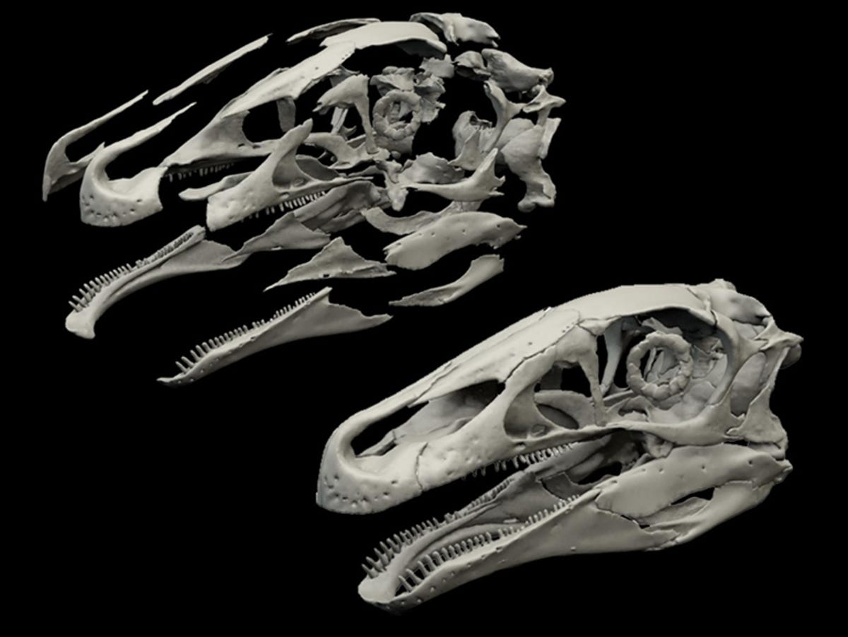
The skull used was that of a 3-4 metre (10-13ft) therizinosaur called Erlikosaurus andrewsi, a herbivorous animal that lived in the Mongolia, 90 million years ago during the Cretaceous Period. As the best preserved and most complete skull of its species, it's highly valuable -- yet even this skull showed obvious damage.
The team scanned the skull, creating a high-resolution 3D model. This model was then virtually disassembled into individual elements, which in turn allowed the team to fill in cracks and breaks, duplicate missing elements, and remove deformations by digitally reversing the steps of that deformation.
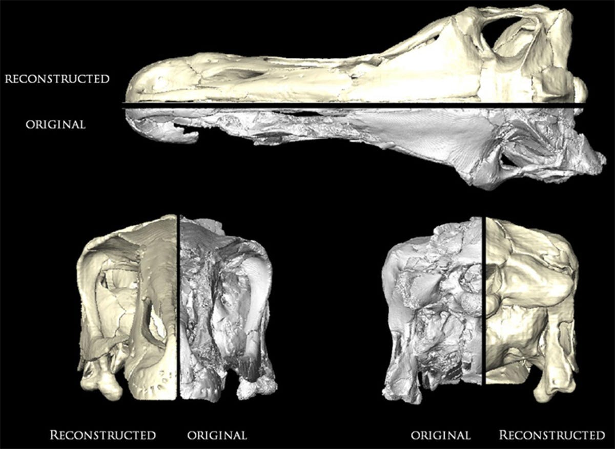
The final step was reassembling the reconstructed elements, resulting in a digital model of a perfect Erlikosaurus skull that can be pulled apart and put back together for study and research without worrying about damaging the fossil.
"Digital models allow the study of the external and internal features of a fossil," co-author Dr Lawrence Witmer said. "Furthermore, they can be shared quickly amongst researchers - without any risk to the actual fossil and without having to travel hundreds or even thousands of miles to see the original."
The team consisted of Dr Stephan Lautenschlager and Professor Emily Rayfield from the University of Bristol, Professor Lindsay Zanno from the North Carolina Museum of Natural Sciences and North Carolina State University, Dr Perle Altangerel from the National University of Ulaanbaatar and Professor Lawrence Witmer from Ohio University. Their full paper, "Cranial anatomy of Erlikosaurus andrewsi (Dinosauria, Therizinosauria): new insights based on digital reconstruction", can be found online in the Journal of Vertebrate Paleontology.