Beautiful art made from bacteria, rat liver cells
An artist and a scientist team up to make striking patterns, self-portraits, and other paintings using a most unlikely medium, "tiny, tiny living things."


Artist Vik Muniz likes working with the super small. One of his recent projects involved etching intricate drawings of sandcastles onto the sides of grains of sand. Now, he's used bacteria and human cells to create striking works of art.
Muniz's work is part of "The Creator's Project," a joint venture between Vice magazine and Intel that "celebrates visionary artists across multiple disciplines who are using technology in innovative ways to push the boundaries of creative expression," according to the project's website.
For his latest art series, "Colonies," the Brazilian multidisciplinary artist teamed with Tal Danino, a postdoctoral fellow at MIT who researches the use of bacteria to help diagnose and treat cancer.
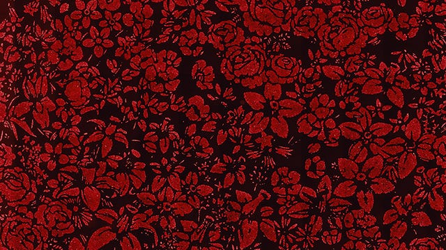

"I always love to collaborate with scientists because it gives me an idea that we can perhaps meet in the middle somewhere and make this perfect piece of art that's exactly this combination between matter and meaning," Muniz told the Creators Project. "'Colonies' is a collaboration between a scientist and an artist, trying to make pictures out of living things -- tiny, tiny living things."
The two came up with a unique three-part process to create images from living cells. Danino described it to The Creators Project as follows:
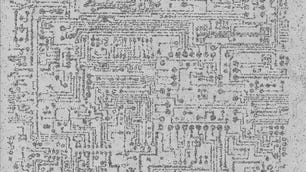
"The process for making these images consists of three steps. The first step is to make a master image wafer using photolithography techniques. The second step is actually to cast a rubber stamp from this wafer, and use that stamp to make a sticky image on a dish that cancer cells or bacteria can stick to. The final step is to apply cancer cells or bacteria to this dish, and allow them to bind to the sticky material, and then take images on a microscope. You can actually see the individual bacteria and cancer cells with this high-powered microscope. But then if you zoom out, you can actually see the image in its entirety."
The "Colonies" series has been exhibited in New York, Tel Aviv, and Sao Paulo, with proceeds from the shows going toward cancer research.
The video above explains the creation process in greater detail, while this gallery will take you through some of the most striking images created in the series.