3D video of sperm could help docs spot strong swimmers
By generating a progressive series of holograms, scientists can watch sperm move and look for structural anomalies that make them less viable, helping to improve odds during in vitro fertilization.


For parents-to-be undergoing in vitro fertilization (IVF) -- the process of joining an egg and a sperm in a petri dish -- using the best sperm of the batch is key to increasing the odds of fertilizing the egg, not to mention getting the job done more efficiently and less expensively. And while today's top tech, called computer-assisted sperm analysis (CASA), provides pretty good imagery, the tracking and imaging system is in 2D.
Now, report researchers from Italy and Belgium in the journal Biomedical Optics Express, a new technique for analyzing sperm does so in 3D and over time, allowing for a far clearer view of sperm concentration and motility (i.e. movement).
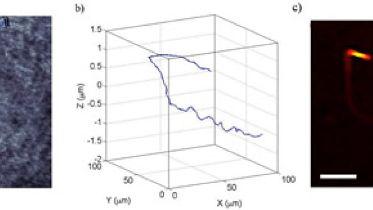
To accomplish this, the researchers combined microscopy with holography (for creating the images in 3D) to visualize sperm not just in the x and y positions, but also according to their depth (z position). And, because they're able to capture video of sperm movement in 3D, "we add a fourth dimension -- time," lead author Giuseppe Di Caprio, who works at the National Research Council in Italy and Harvard University, said in The Optical Society news release.
The researchers say they first had to separate laser light into two beams so that they could transmit one through a petri dish with swimming sperm cells, then magnify the image of the swimmers with a microscope, and finally add the second, superimposed beam that provides an interference pattern they record on video. What they get, then, is a progressive series of holograms that lets them watch sperm move and look for any structural anomalies.
Di Caprio says their approach is called digital holographic microscopy (DHM), and that while the data they collected on sperm motility and shape is consistent with what has been gathered in other studies, they now have a far better view of cause and effect between these two characteristics.
"For example, we found that most of the sperm cells we observed swim along in one plane as expected. However, with the more detailed analysis provided by DHM, we also were able to show that this 'in-plane' movement -- which we believe is linked to higher potential for fertility -- does not occur when there are morphological anomalies such as sperm with misshapen heads or 'bent tails.'"
Ultimately, this new tracking tech may let scientists use microchips to do what women surely wish they could do via pure intuition: sort through the big batch and find the most viable contenders.