The Art of Science draws beauty from medical research
The exhibition features artwork created from scans of cancer cells and protein.


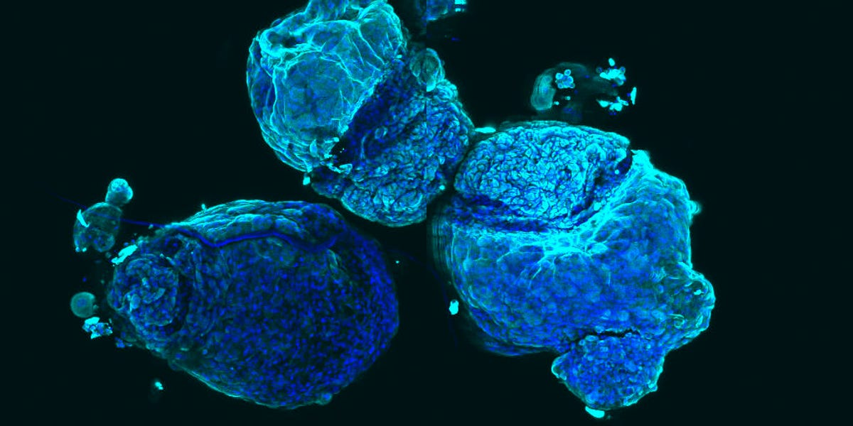
'Croissant' by Casey Ah Cann, ACRF Stem Cells and Cancer division
Here you see lung stem cells that were grown in a lab in the form of a pneumosphere. The red-colored nucleus of each cell is surrounded by tublin, covered in blue.
"In this experiment, I was able to disrupt the function of a specific gene in these cells and see if that changed the structure of the individual cells or of the whole pneumosphere shape," said Ah.
"The specific pneumosphere presented here was a control, or an unaltered set of cells/pneumosphere. With this I was able to compare and contrast the normal cells with my disrupted cells. These kinds of techniques are so important to our understanding of biology. Only by understanding things in their normal state can we start to tease apart disease and develop better treatments."
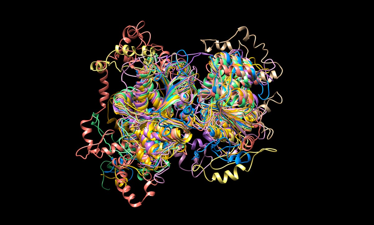
'The gobstopper' by Brendan Ansell, Balu Balan, Aaron Jex, Population Health and Immunity division
This computer simulated image shows the proteins of the parasite Giardia, which infects humans after it is ingested. It is particularly dangerous in young children, and treatment for the parasite is "both unpleasant and not completely effective," according to the exhibition caption.
"Through producing and studying images of protein structure, we can identify regions of similarity in these proteins that would allow us to make an effective drug to jam up the parasite's biology. This would allow us to stop the transmission of disease. Giardia is the most common human parasite, causing acute diarrhoea in both developing and developed countries."
'Cnidaria' by Clare Weeden, ACRF Stem Cells and Cancer division
Another look at our lungs: the airway stem cells from a lung cancer patient. The light blue color is the lung protein that provides structure to the airways.
"The dark blue colour marks the nuclei of each cell, whilst the light blue colour is lung protein that provides structure to our airways. By identifying and culturing human lung stem cells, we can develop a broader understanding of how the lung functions normally and how these processes go awry in lung cancer."
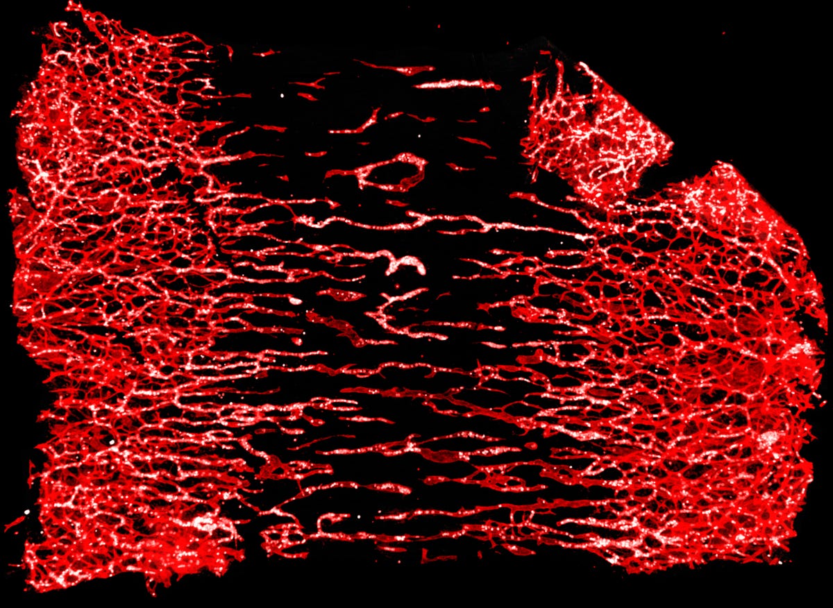
'Meet halfway' by Alison Farley, Molecular Medicine division
These are blood vessels and white blood cells migrating from the side of a mouse embyro skin. This was an experiment on the role of platelets in blood vessel development.
"This work is important in understanding when and why brain bleeds/stroke occurs in the absence of platelets."
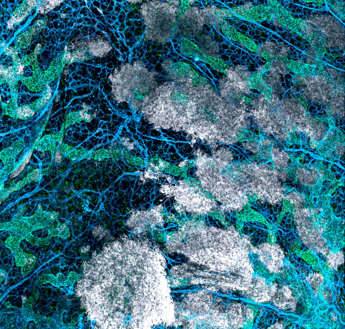
'Bird's eye view' by Alison Farley, Molecular Medicine division
Here are blood vessels again in blue alongside green lymphatic vessels and white blood cells leaking from a mouse with no platelets. The scientists are investigating "the role of platelets in vasculature development in the embryo and the role of platelets in lymphatic and blood vessel separation."

'M-Galaxy' by Roberto Bonelli, Population Health and Immunity division
Looking very much like a star field, the image you see represents the 23,621 connections between the 868 metabolites (a substance needed for metabolism) measure in the blood of healthy people and patients affected by Macular Telangiectasia, a rare eye disease.
Blue lines are positive connections, while red are negative, with the outer ring comprising of metabolites with few or no connections. As no one knows what yet causes the disease, scientists are hoping that metabolism links here could contain an answer -- apparently groups composed by big metabolites were important to the eye disease.
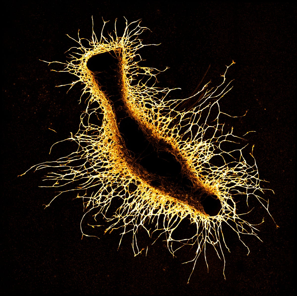
'Creature of the deep' by Zoe Grant, Development and Cancer division
While it looks very much like a fish, it's actually blood vessels growing out of a small bone piece in a laboratory dish. The scientists watch the blood vessels grow to understand how it's controlled.
Understanding how it works is important, as scientists can then figure how blood vessel growth contributes to diseases such as cancer.
"We want to understand how blood vessels grow so we can try to block their growth in these diseases."
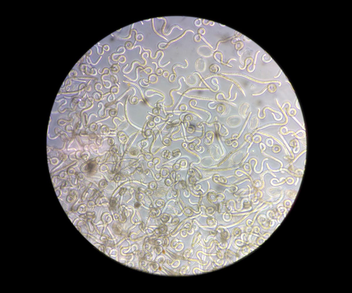
'Ascaris kindergarten' by Louise Baker, Population Health and Immunity division
If you hate worms, you should probably not look any closer. These are Ascaris suum larvae, which tend to live in the soil and are often found in developing countries causing "significant gastro-intestinal disease and chronic malnutrition and developmental stunting in children."
Scientists are hoping to figure out how these nematodes infect and develop within new hosts and how to treat infections.
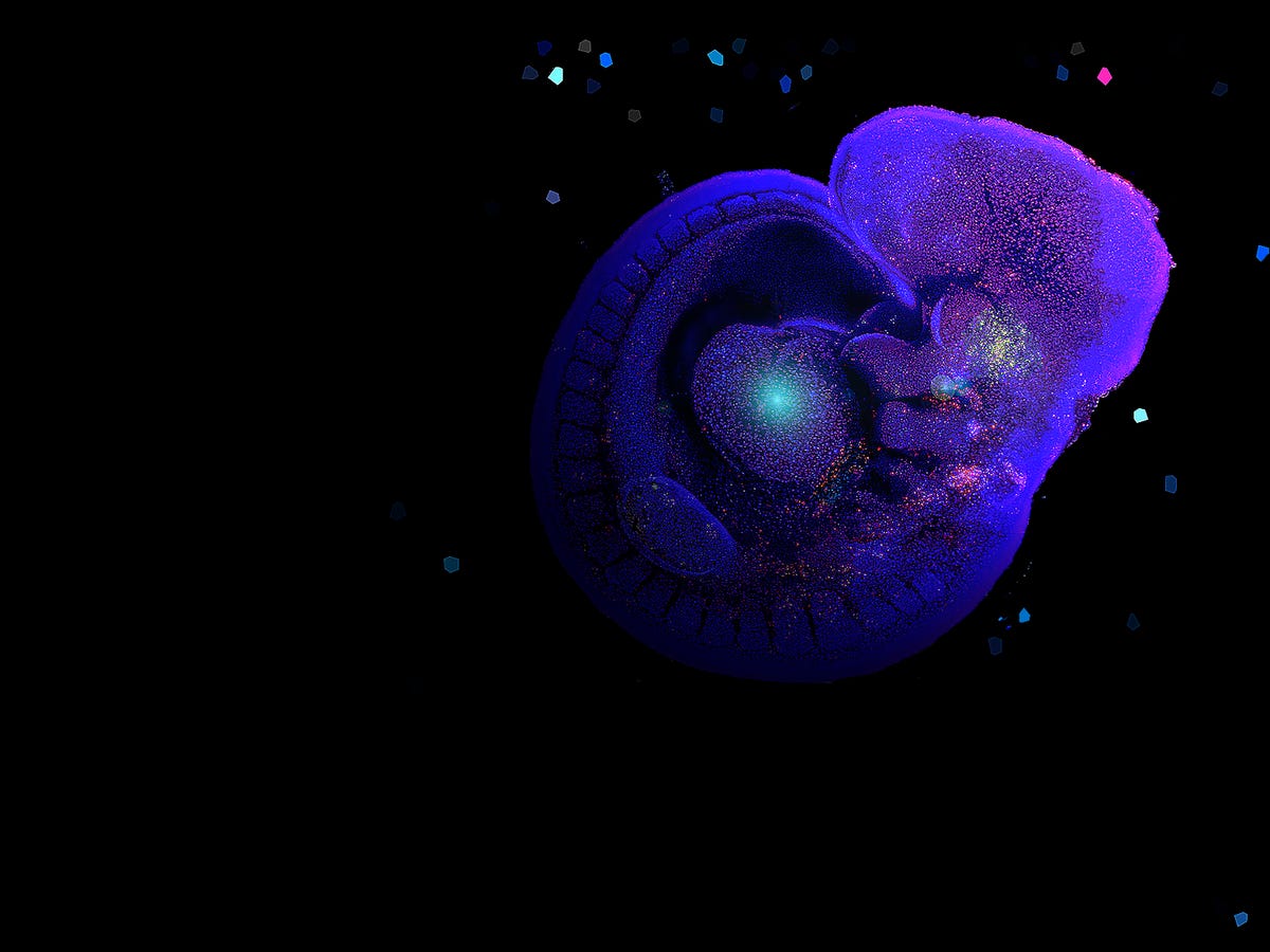
'Starry starry night' by Francine Ke, Molecular Genetics of Cancer division
While it may look like a scene from a sci-fi movie in space, these "stars" are actually a cell in an embryo. The brightly stained cells are dying, a process called apoptosis, while the dark blue cells are still alive.
"My research focuses on understanding the interplay between apoptosis and embryonic development, and how a lack of cell death or too much cell death can lead to physical abnormalities," said Ke.
"For instance, do you know that our fingers and toes first develop as webs, and apoptosis subsequently occurs to give us individual digits? On a similar note, defects such as spina bifida and cleft palate are also caused by disrupting the fine balance between cell survival and death. It is important to learn about these processes so that we can help decrease the occurrence of such abnormalities in future."

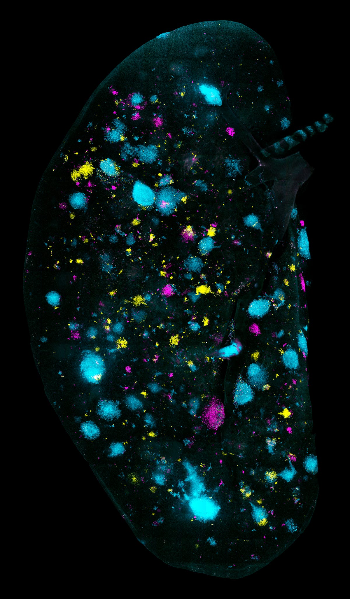
'Electrifying' by Raymond Yip, ACRF Stem Cells and Cancer division
This amazing picture is collected from mice with aggressive breast tumors, created using 3D imaging. The micro vessels in cyan and yellow wrap the ball of magenta tumor cells -- and seem to indicate that tumor cells induce new blood vessels to help them grow within the bone marrow.
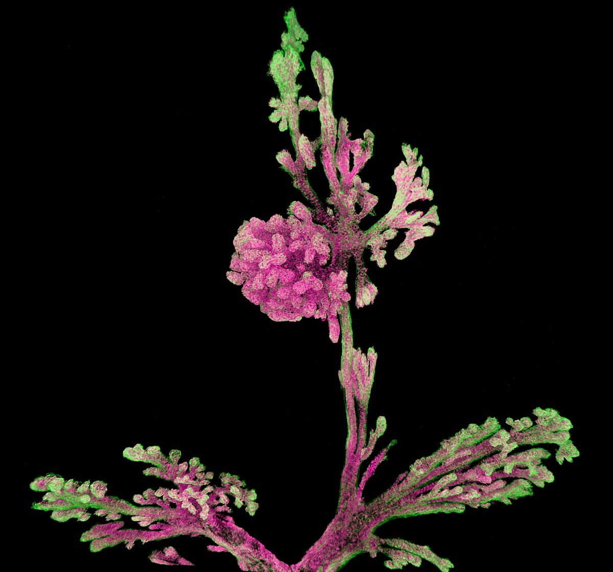
'Blossom' by Bianca Capaldo, ACRF Stem Cells and Cancer division
This flower-like picture is actually the "terminal lobular alvolar" found at the end of mammary ducts. Scientists are studying the architecture and cellular composition of these "flowers" as breast cancer usually starts here.
'Splatter painted lung' by Caleb Dawson, ACRF Stem Cells and Cancer division
This colorful image is actually quite a scary picture of a lung covered in multi-colored tumors. Scientists specifically grew these colorful tumors and introduced them to a lung to track how cancer escapes and spreads.
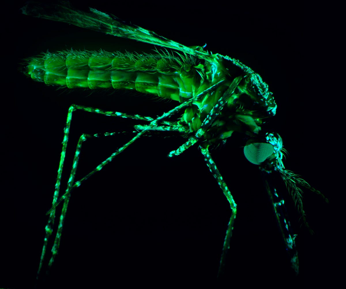
'Humanity's deadliest foe' by Qike Wang, Molecular Medicine division and Julie Healer, Infection & Immunity division
Here's something most of us hate -- a mosquito transmitting malaria. These mosquitoes are bred in an insectary and then infected with the malaria parasite to help scientists discover tools to stop the disease.
More Galleries

My Favorite Shots From the Galaxy S24 Ultra's Camera
20 Photos
Honor's Magic V2 Foldable Is Lighter Than Samsung's Galaxy S24 Ultra
10 Photos
The Samsung Galaxy S24 and S24 Plus Looks Sweet in Aluminum
23 Photos
Samsung's Galaxy S24 Ultra Now Has a Titanium Design
23 Photos
I Took 600+ Photos With the iPhone 15 Pro and Pro Max. Look at My Favorites
34 Photos