Turning a regular microscope into billion-pixel imaging system
Engineers at Caltech say that their approach -- computing their way past optical limitations -- could bring high-performance microscopes to medical clinics in developing countries.


Ah, physics. The cold, hard reality of how things work so often gets in the way of how we would prefer them to work.
But when it comes to the field of microscopy, which has been held back by the physical limitations of optical lenses, a group of engineers at Caltech say they've been able to use a computational approach to bypass these limitations -- and that the final images produced using their new system contain 100 times more information. What's more, the system costs just $200 to implement with a conventional microscope.
In a nutshell, the limitations of optical lenses have forced researchers to pick and choose between a system that gives them high resolution over a small field of view or low resolution over a wider field of view. See a little bit clearly or a lot coarsely.
"We found a way to actually have the best of both worlds," Guoan Zheng, lead author of the new paper in Nature Photonics, said in a school news release. "The optical performance of the objective lens is rendered almost irrelevant, as we can improve the resolution and correct for aberrations computationally."
Take that, physics!
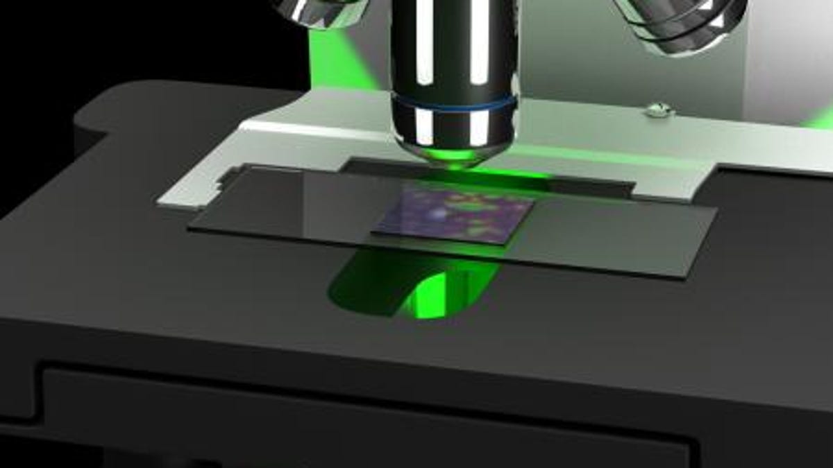
Ultimately, the researchers were able to improve the resolution of a conventional 2X objective lens to the level of a 20X -- so they combined the larger field-of-view advantage of the 2X lens with the resolution of the 20X lens -- for just $200. They achieved this by taking 150 low-res images of a sample, corresponding each image to an LED element in an LED array, and then using a novel computational approach called Fourier ptychographic microscopy, stitching together the low-res images to form high-res intensity, resulting in a far more complete picture of what is available across the entire light field of the sample.
Changhuei Yang, professor of electrical engineering, bioengineering and medical engineering at Caltech, says that we can only sense variations in intensity when looking at light from an object, but that light actually varies not only in intensity but in its phase -- and that phase is related to the angle at which light travels. By teasing out both the intensity and the phase of the light field across low-res images, they are able to "correct for optical aberration issues that otherwise confound your ability to resolve objects well."
Because the approach does not require scanning various parts of an image -- an entire sample can be imaged all at once via these 150 separate points -- and because the system gathers all the data about the light field, it is not only a faster approach, but it could actually correct errors computationally, such as blurriness, so that clinicians do not need to rescan an image for digital pathology applications.
"One big advantage of this new approach is the hardware compatibility," Zheng added. "You only need to add an LED array to an existing microscope. No other hardware modification is needed. The rest of the job is done by the computer."
The researchers figure their system could have many applications in anything from digital pathology to forensic photography, and that it could even extend to other imaging techniques, such as X-ray and electron microscopy.