Sharpest ever images of living bacteria reveal unexpected membrane structure
The images are so clear that scientists can see proteins on a bacterium's outer membrane.


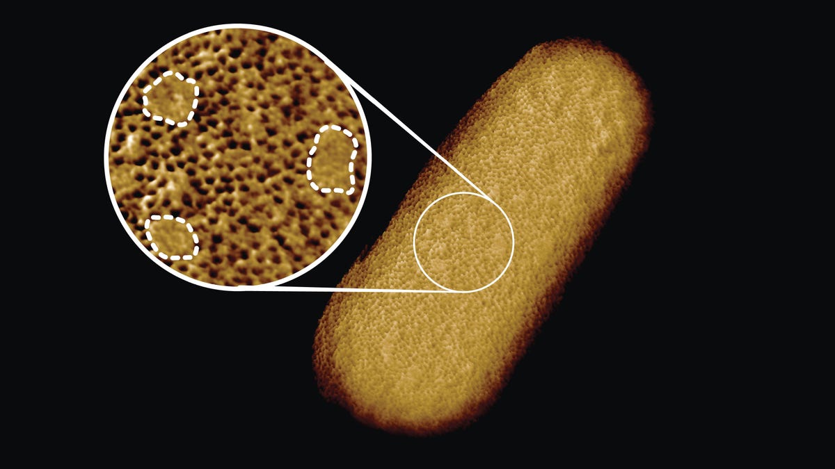
This image of a live Escherichia Coli bacterium reveals the structure of its outer membrane. In the inset image, the dotted lines signify regions of the membrane that lack proteins.
Using a microscopy technique that pulls back the curtain on the nanoscale world, researchers have generated the sharpest images of live bacteria ever taken. The high-resolution snaps reveal a patchwork of proteins that make up the outer membrane and, potentially, give scientists a new way to attack the organisms.
In a study published Monday in the journal Proceedings of the National Academy of Sciences, researchers from the US and UK provide images of the rod-shaped bacterium Escherichia Coli, which is commonly found in our gastrointestinal tracts and helps digest food. It's a mostly harmless citizen of our guts, but certain E. Coli strains can cause severe illness. In recent years, scientists have shown that these strains are becoming more resistant to antibiotics.
The bacteria are, of course, incredibly tiny. About 150 to 300 individual bacteria could probably fit head-to-toe across the period at the end of this sentence. While regular microscopes can see the bacteria as clumps of rods scattered all across a glass slide, they don't give us a good idea of the structure of a bacterium's body and the outer membrane that protects it from our drugs.
"The outer membrane is a formidable barrier against antibiotics and is an important factor in making infectious bacteria resistant to medical treatment," Bart Hoogenboom, a nanotechnologist at University College London and co-author on the paper, said in a press release. Hoogenboom and his team wanted to take a much closer look and turned to "atomic force microscopy" to study the membrane in greater detail.
Atomic Force Microscopy, or AFM, is a powerful technique to study objects that are just nanometers in length. (A nanometer is a millionth of a millimeter.) Hooked up to a computer, the device can study the surface of tiny, tiny objects by running a fine-tipped probe, just a few nanometers wide, across the surface. The probe moves along the object -- whether it's a microbe, a piece of bone or another nanomaterial -- and can sense the undulations present. You can imagine it kind of like a turntable stylus, moving along the grooves of a vinyl record. Totally different process but similar concept.
When the team ran the AFM probe over the surface of a living E. Coli bacterium, they discovered it was a mosaic of proteins. Most areas of the outer membrane were covered in mostly immobile proteins, but there were also islands that lacked any protein at all and were instead full of sugary lipid molecules.
"This suggests that the barrier may not be equally hard to breach or stretch all over the bacterium, but may have stronger and weaker spots that can also be targeted by antibiotics," Hoogenboom said.
Outer membranes are present on what's known as "gram-negative" bacteria, like E coli, and not gram-positive bacteria, like Staphylococcus. The "gram" doesn't refer to weight, but a specific test designed to quickly differentiate bacteria based on their cell walls. Notably, gram-negative bacteria have a more impenetrable cell wall, which makes it hard for antibiotics to get in and work to destroy the microbes.
Antibiotic resistance makes infectious diseases harder to treat and can turn common infections into life-threatening scourges. Many antibiotic resistant infections are picked up in hospitals, and the World Health Organization has consistently listed antibiotic resistance as a global health threat over the past few years because it has the potential to upend health care systems.
How the protein patchwork of the outer membrane might affect the strength of a bacteria's protective covering or how it could help combat antibiotic resistance is not yet known. But with advanced microscopy techniques, scientists can shine a light on a previously unseen realm.