X

IBM microfluidics tech designed to improve cancer diagnosis

A technique to pump tiny amounts of marker fluids onto a test sample could enable many more tests during a biopsy and therefore a better understanding of a person's cancer.

Stephen Shankland Former Principal Writer
Stephen Shankland worked at CNET from 1998 to 2024 and wrote about processors, digital photography, AI, quantum computing, computer science, materials science, supercomputers, drones, browsers, 3D printing, USB, and new computing technology in general. He has a soft spot in his heart for standards groups and I/O interfaces. His first big scoop was about radioactive cat poop.
Expertise Processors, semiconductors, web browsers, quantum computing, supercomputers, AI, 3D printing, drones, computer science, physics, programming, materials science, USB, UWB, Android, digital photography, science. Credentials
  • Shankland covered the tech industry for more than 25 years and was a science writer for five years before that. He has deep expertise in microprocessors, digital photography, computer hardware and software, internet standards, web technology, and more.
Stephen Shankland
2 min read
IBM's device for speeding up the identification of cancer cells pumps a tiny amount of fluid through a very small tip into a biopsy sample on a microscope. The tip then sucks the fluid back out again so it doesn't stain more than a tiny patch.
IBM's device for speeding up the identification of cancer cells pumps a tiny amount of fluid through a very small tip into a biopsy sample on a microscope. The tip then sucks the fluid back out again so it doesn't stain more than a tiny patch. Stephen Shankland/CNET

ZURICH -- Researchers accustomed to designing tiny features on microprocessors have taken up a new tiny-technology challenge: improving the diagnostic tests used to spot cancer.

Using a procedure called a biopsy, pathologists today closely examine cells to try to determine if a person has cancer and if so, details about what type. Such tests use chemical markers that can spotlight a variety of problems, including different types of cancer, but the tiny slice that constitutes a biopsy sample isn't big enough for a multitude of tests.

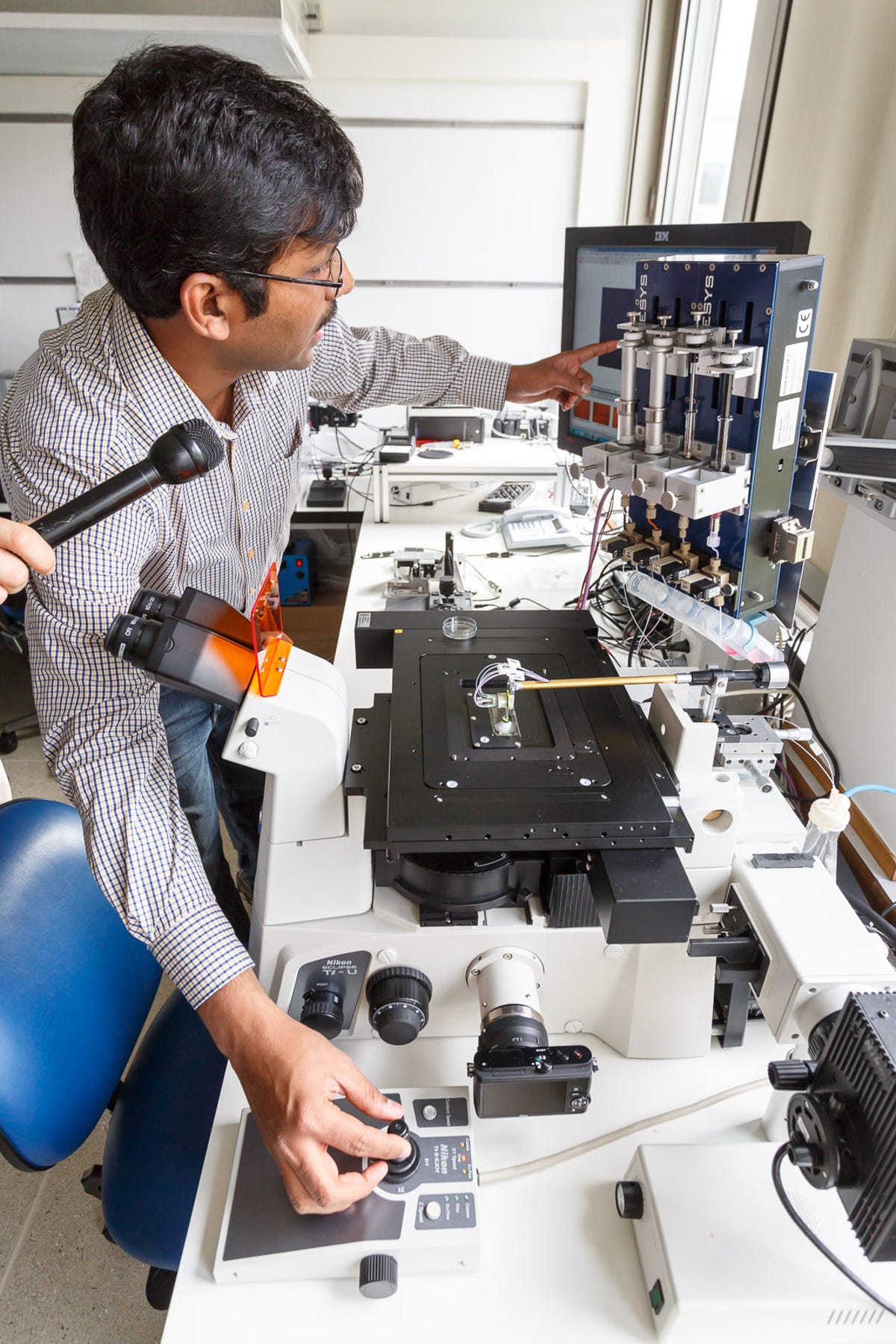
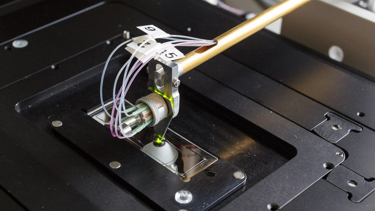
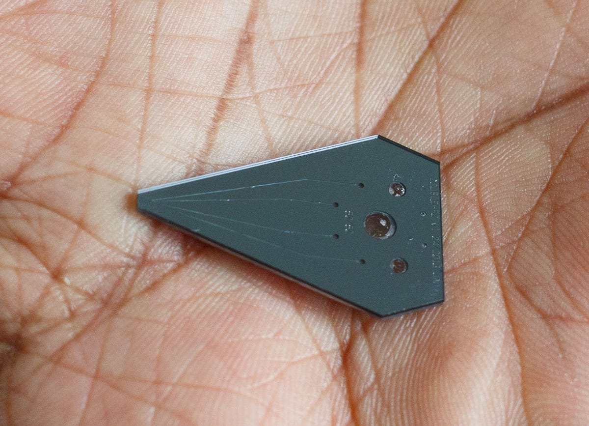
IBM's approach, which University Hospital Zurich plans to test in the coming months, uses a chip technology called microfluidics to shrink the area required for such tests to a square patch just 100 micrometers wide -- about the same as a human hair. It pumps the marker chemical down one tiny channel into the biopsy sample, then slurps it back up with a second channel to keep the marker from spreading beyond the confines of its designated patch, and a pathologist watches on a microscope to see the response.

This silicon-based "microfluidics" device channels tiny amounts of special liquids onto a biopsy specimen so pathologists can identify different forms of cancer.
This silicon-based "microfluidics" device channels tiny amounts of special liquids onto a biopsy specimen so pathologists can identify different forms of cancer. Stephen Shankland/CNET

"It's a very economical way of using critical tissue samples," said Emmanuel Delamarche, an IBM Research Zurich expert in nanotechnology matters such as self-assembling devices. A typical sample measuring 1 square centimeter has room for 500 test patches on just 5 percent of its surface, he said.

With the technique today, a pathologist can steer the tip around with a joystick so it puts down the marker in just the right area; the channels in the tip itself are just a few millonths of a meter wide. With several reservoirs of markers attached, the pathologist can switch among several markers.

Researcher Emmanuel Delamarche, from IBM's Zurich labs explains the company's technique for speeding up the identification of cancer cells.
Researcher Emmanuel Delamarche, from IBM's Zurich labs explains the company's technique for speeding up the identification of cancer cells. Stephen Shankland/CNET

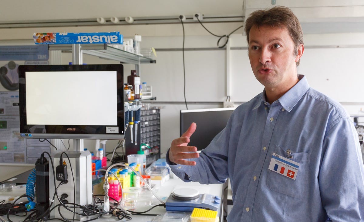
In the initial incarnation, a pathologist watches the results on a display. But Delamarche hopes eventually the system could work much more automatically: A sample is inserted into a box and test results emerge shortly afterward without human intervention.

"We need to partner to re-engineer our technology to make it into an instrument that is very reliable," Delamarche said.

Govind Kaigala from IBM Research in Zurich demonstrates the microfluidics technology mounted atop an inverted microscope (one that looks upward). He positions the device, attached to the end of the brass-colored stalk, over a slide containing a biopsy specimen. A joystick in his right hand changes position, and with his left he points to reservoirs of the marker chemicals used to identify cancer cells.
Govind Kaigala from IBM Research in Zurich demonstrates the microfluidics technology mounted atop an inverted microscope (one that looks upward). He positions the device, attached to the end of the brass-colored stalk, over a slide containing a biopsy specimen. A joystick in his right hand changes position, and with his left he points to reservoirs of the marker chemicals used to identify cancer cells. Stephen Shankland/CNET