How to better spy on a childhood virus
Scientists at Georgia Tech say a new technique for tagging the genome and studying the RNA of a virus could help them discover better antiviral drugs and perhaps even more effective vaccines.


Influenza, Ebola, and respiratory syncytial viruses (RSV) can be nasty little buggers, infecting their hosts with rash abandon and, especially when they attack young babies, even killing them. And the danger reaches beyond the very young. Pneumonia, for instance, is the leading cause of death in children worldwide, according to the World Health Organization, and RSV is the most common viral cause of pneumonia.
As imaging techniques advance, researchers are being able to study these viruses in greater and greater detail. Now, according to a team of scientists at Georgia Tech, Vanderbilt, and Emory, one new technique for studying RSV in microscopic detail could help them spy on the structure of the virus for days and help them better understand how it enters cells, how it replicates, why some lung cells manage to escape the wrath of the virus, and so on. These discoveries, in turn, could pave the way for better antiviral drugs and even a vaccine to prevent infection in the first place -- and the scientists say there's no reason the approach can't extend to other viruses as well, including influenza and Ebola.
At the heart of the technique, which is described in the journal ACS Nano, is the ability to tag the virus genome with a probe that does not change the behavior of the virus but still allows researchers to follow and study it for days.
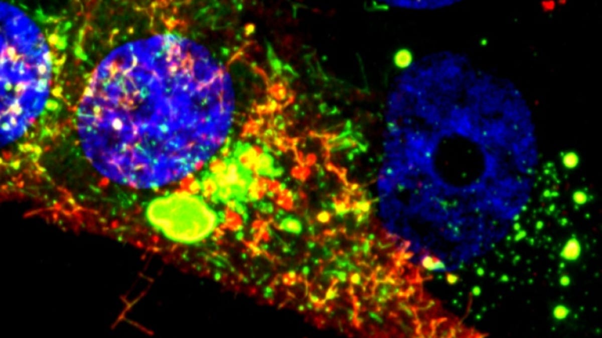
To do this, the researchers turned to a method they developed a few years ago for labeling RNA viruses that uses what they call multiply-labeled tetravalent RNA imaging probes (MTRIPS). These little guys are complex, able to evade cellular defenses, and latch tightly onto the virus' RNA without actually changing the behavior of the virus. They use multiple fluorophores to highlight the viral RNA for easier visual tracking (thanks to standard microscopy tech) as it enters host cells and as infectious particles exit those cells and spread.
To make all of this possible, graduate student Eric Alonas also had to concentrate the RSV without changing its ability to infect, which in turn could have changed its ability to enter host cells. In a school news release, biomedical engineering associate professor Philip Santangelo said it took a lot of work to concentrate the virus, but now that they got the right techniques down, "we can make lots of infectious virus that's labeled and can be stored so we can use it when we want to."
Now that they can store the virus and spy on it at nanoscale resolution for days, Santangelo said the next step is to figure out why some cells are "exploding with virus" while others seem somehow immune. "If you look at a field of cells, you see huge differences from cell to cell, and that is something that's not understood at all...Perhaps we can figure out a way to help the bad ones look more like the good ones."
Doing that may lead to not only better drugs to treat infection but possibly even an RSV vaccine to prevent it altogether.