3D-printed cadavers revolutionise anatomical education
The 3D Printed Anatomy Series allows medical students to learn human anatomy without needing access to a real cadaver.


Since ancient history, humans have been dissecting the cadavers of our own kind to understand how the body works. The ancient Egyptians, who dissected and mummified their dead, had medical skills far beyond the level of the rest of the world at that time. In ancient Rome, the famous medical researcher Galen, forbidden from dissection human corpses, used pigs and primates to gain an understanding of human anatomy. The history of surgical research is riddled with grave-robbers and clandestine operations.
It's easier now in many parts of the world, but it can be dependent on a variety of factors; whether or not the deceased has given permission for their body to be used for the purposes of research for example. And in other regions of the world, the use of real cadavers can still be problematic.
The 3D Printed Anatomy Series, created by researchers at Australia's Monash University, offers a solution. The kit consists of all the major body parts required to learn the anatomy of the limbs, chest, abdomen, head and neck -- all without containing any actual human body parts.
"For centuries cadavers bequested to medical schools have been used to teach students about human anatomy, a practice that continues today. However, many medical schools report either a shortage of cadavers, or find their handling and storage too expensive as a result of strict regulations governing where cadavers can be dissected," explained Professor Paul McMenamin, Director of the University's Centre for Human Anatomy Education.
"Without the ability to look inside the body and see the muscles, tendons, ligaments, and blood vessels, it's incredibly hard for students to understand human anatomy. We believe our version, which looks just like the real thing, will make a huge difference."
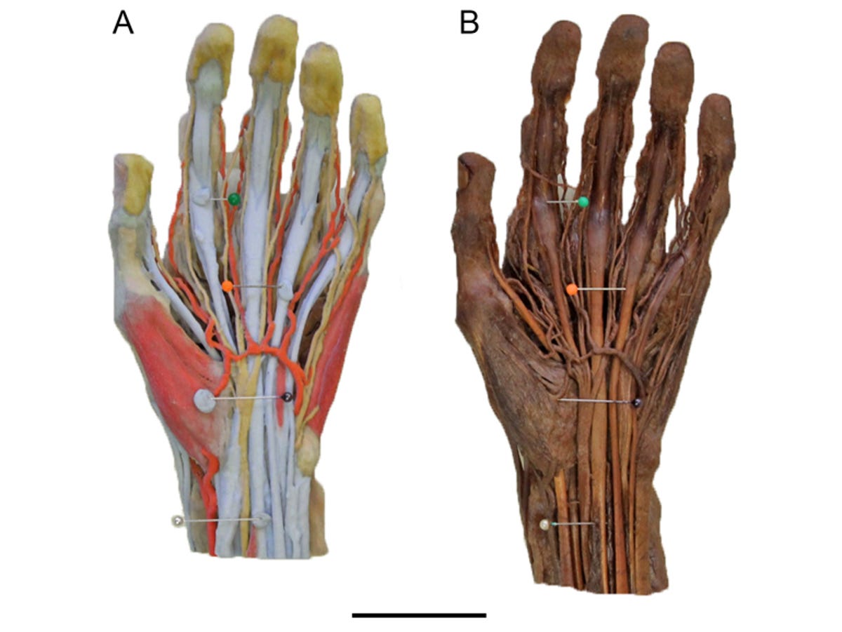
The 3D Printed Anatomy Series is created from real humans. First, the team performed scans, either X-ray CT scans or surface scans. These scans are then used to create a printable 3D model of the body parts, which are then sent to a high-resolution 3D printer and printed either in full colour in a plaster-like powder or plastic.
"Radiographic imaging, such as CT, is a really sophisticated means of capturing information in very thin layers, almost like the pages of a book," Professor McMenamin said. "By taking this data and making a 3D rendered model we can then colour that model and convert that to a file format that the 3D printer uses to recreate, layer by layer, a three-dimensional body part to scale."
More information about the project, which is currently seeking commercial partners, can be read online in the journal Anatomical Sciences Education.