The incredible tiny world of microbiology
The winners of Olympus' annual live sciences photography competition are in, with the top 10 submissions revealing an entire world of microscopic wonder.


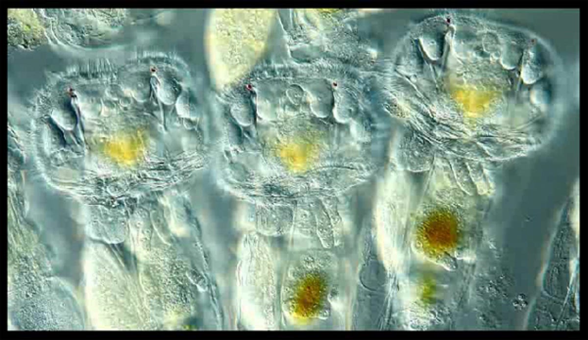
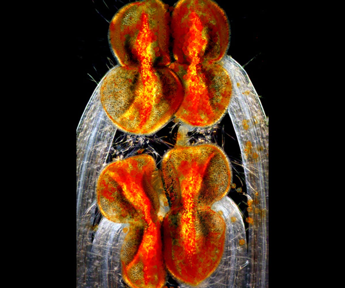
Colonial rotifers showing eyespots and corona, magnification 200x to 500x
The winners of Olympus' annual live sciences photography competition are in, with the top 10 submissions revealing an entire world of microscopic wonder.
It's the 10th year of Olympus' BioScapes international digital-imaging competition — where photographers from around the globe can send in their photos and videos of a world that usually goes unseen by human eyes. Captured via light microscope, the images are of live science specimens studied in laboratories — and entrants can use any magnification, lighting and brand of equipment.
Hidenao Tsuchiya, Olympus America's vice president and general manager for the Scientific Equipment Group, said of the competition:
Microscope images forge an extraordinary bond between science and art. We founded this competition to focus on the fascinating stories coming out of today's life science research laboratories. The thousands of images that people have shared with the competition over the years reflect some of the most exciting work going on in research today — work that can help shed light on the living universe and ultimately save lives. We look at BioScapes and these beautiful images as sources of education and inspiration to us and the world.
This year's winner was Ralph Grimm, a teacher from Jimboomba, Queensland, a small town about 45 kilometres south of Brisbane. Grimm's work in microbial imaging has won multiple awards and accolades in the past, including honourable mentions and "image of distinction" awards in this year's Nikon Small World competition. His winning BioScapes entry is a 58-second video — the first video to win the competition — capturing the rapid movements of pond-dwelling colonial rotifers' cilia, with their red eyes clearly visible.
We've collected the top 10 winners here for your viewing pleasure (in order of placement), but you can check out the full gallery on the official site, which includes an extensive and beautiful collection of honourable mentions.
If you want to enter the 2013 competition, visit the BioScapes website for the terms and conditions.
Ralph Grimm
Jimboomba, Queensland, Australia
Captured using differential interference contrast microscopy.
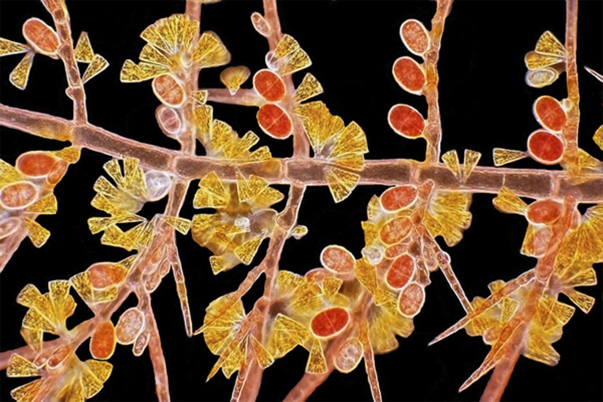
Red algae Scagelia, showing reproductive tetraspores and golden diatoms
Dr Arlene Wechezak
Anacortes, Washington, US
Captured using dark field microscopy.

East-coast US fern, Polypodium virginianum, showing a cluster of spore-filled sporangia and specialised protective hairs called paraphyses
Dr Igor Siwanowicz
HHMI Janelia Farm Research Campus, Ashburn, Virginia, US
Captured using confocal microscopy.
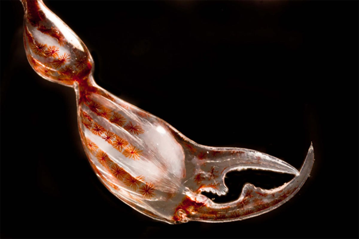
Claw of crustacean amphipode Phronima sp (pram bug — deep sea plankton), showing muscles and rows of pigment cells
Dr Christian Sardet and Sharif Mirshak
The Plankton Chronicles Project, Villefranche-sur-Mer, France, and Montreal, Quebec, Canada
Captured using dark field microscopy.
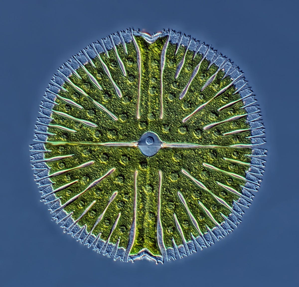
Unicellular green alga Micrasterias from lake sample
Rogelio Moreno Gill
Biomarcell Station Zoologique, Panama City, Panama
Captured using differential interference contrast microscopy, comprising 22 stacked images.
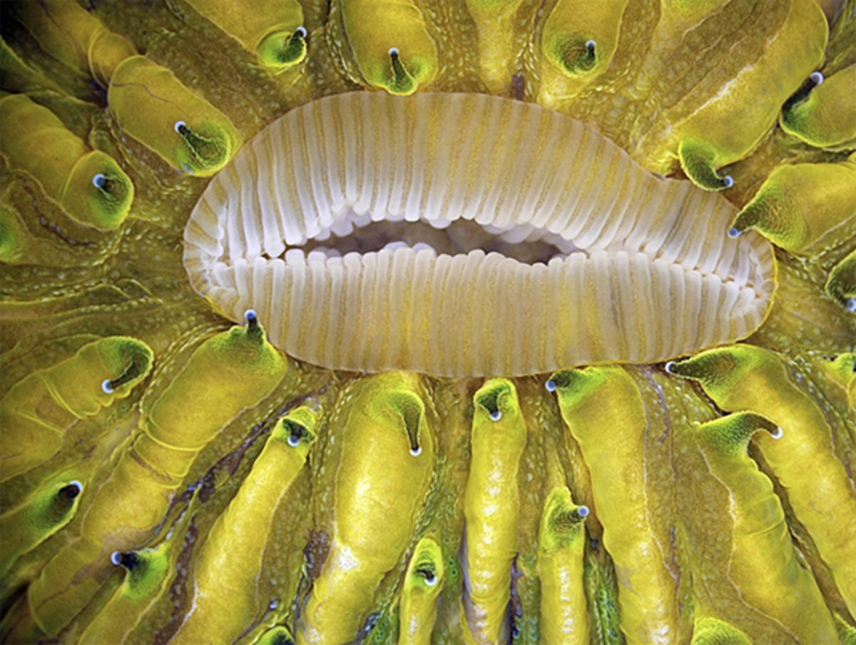
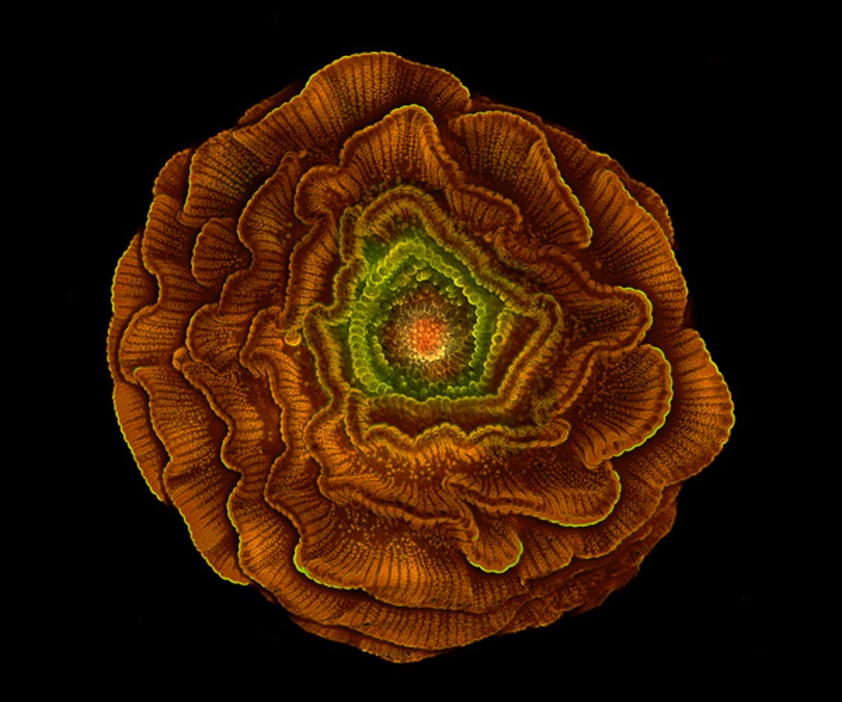
Live mushroom coral Fungia sp — close-up of mouth during expansion
James Nicholson
NOAA/NOS/NCCOS Center for Coastal Environmental Health & Biomolecular Research, Fort Johnson Marine Lab, Charleston, South Carolina, US
Captured using autofluorescence.
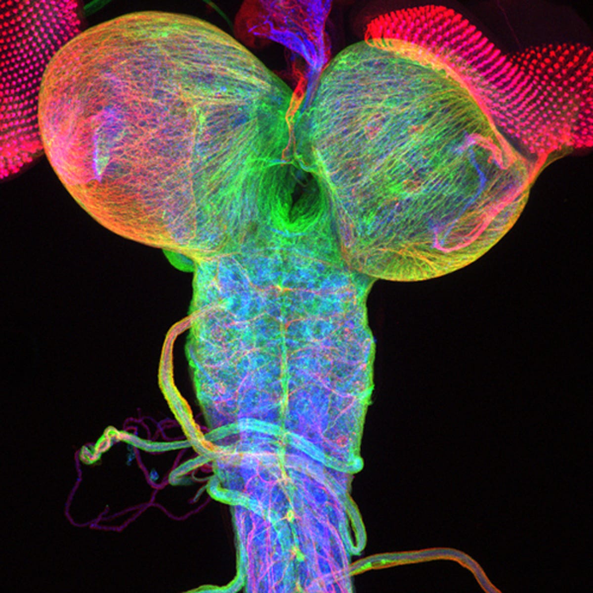
Beta-tubulin expression of a Drosophila third instar larval brain, with attached eye imaginal discs
Dr Christian Klämbt and Dr Imke Schmidt
University of Münster, Münster, Germany
Captured using confocal microscopy.
Henbit (Lamium amplexicaule) stamens anthers and filaments
Edwin Lee
Carrollton, Texas, US
Captured using phase contrast microscopy.
Delphinium seed
Sahar Khodaverdi
University of Tabriz, Tabriz, East Azerbaijan, Iran
Captured using epifluorescence, comprising multiple stacked images.
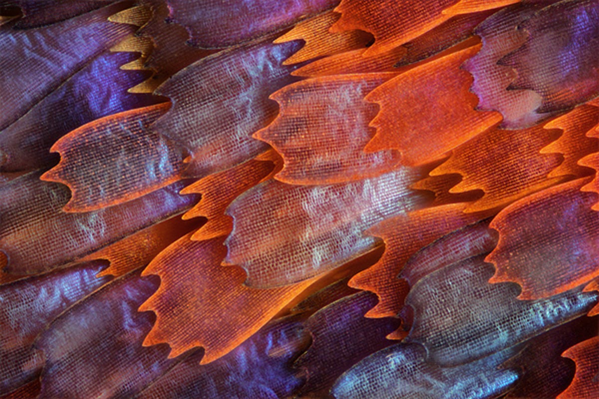
Butterfly "Prola Beauty" (Panacea prola) wing scales, 200x
Charles Krebs
Issaquah, Washington, US
Captured using diffuse reflection illumination.
More Galleries
My Favorite Shots From the Galaxy S24 Ultra's Camera
20 Photos
Honor's Magic V2 Foldable Is Lighter Than Samsung's Galaxy S24 Ultra
10 Photos
The Samsung Galaxy S24 and S24 Plus Looks Sweet in Aluminum
23 Photos
Samsung's Galaxy S24 Ultra Now Has a Titanium Design
23 Photos
I Took 600+ Photos With the iPhone 15 Pro and Pro Max. Look at My Favorites
34 Photos