With X-ray tech, scientists can peer inside cells
Using an imaging technique similar to a CAT scan, researchers are able to build a kind of whole-cell portrait.


- Extensive journalism experience in digital media.
Scientists have developed a way to look inside a whole cell that doesn't involve the usual method of slicing and staining in the lab. Instead, you might say they're employing X-ray vision.
By using soft X-ray tomography (SXT), researchers can take images of a cell every 100 milliseconds and then re-create a whole picture of it from about 90 to 200 images in just a few minutes. The news was presented Friday at the annual meeting of the American Association for the Advancement of Science, according to Science Now. (The AAAS publishes Science Now.)
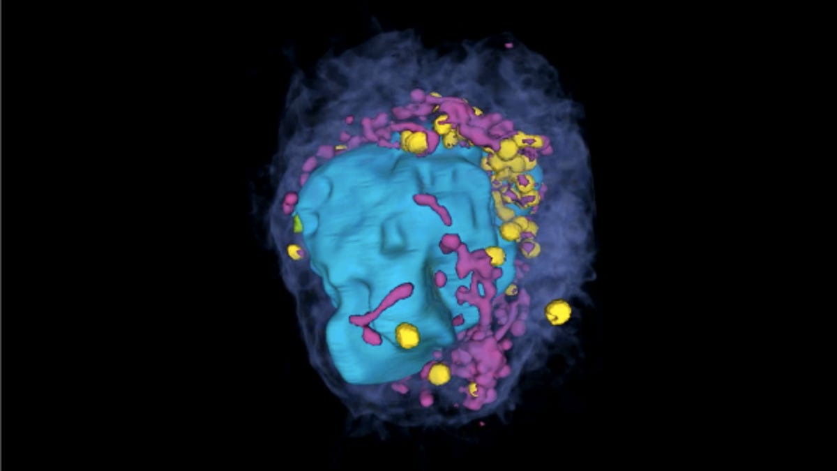
In the image of a T cell above, the scientists have color-coded the lysosomes yellow, the mitochondria pink, and the nucleus blue. Check out the video to see the so-called whole-cell portrait.