Off-the-shelf digital camera sees cancer in real time
Biomedical engineers and university cancer researchers tweak an on-market camera that can distinguish between healthy cells and cancerous ones.


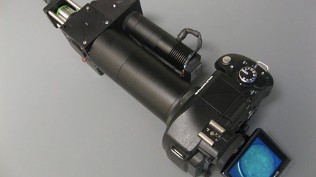
Using a $400 Olympus E-330 digital camera, Rice University biomedical engineers and University of Texas cancer researchers report in PLoS ONE this week that they are able to distinguish between healthy and cancerous cells with only a little tweaking.
"Consumer-grade cameras can serve as powerful platforms for diagnostic imaging," says lead author and Rice professor Rebecca Richards-Kortum in the school's news release. "Based on portability, performance, and cost, you could make a case for using them both to lower health care costs in developed countries and to provide services that simply aren't available in resource-poor countries."
The team captured images of cells using a small bundle of fiber-optic cables attached to the E-330. They then applied a fluorescent dye that makes the nuclei of cells glow brightly when lighted with the tip of the fiber-optic cables.
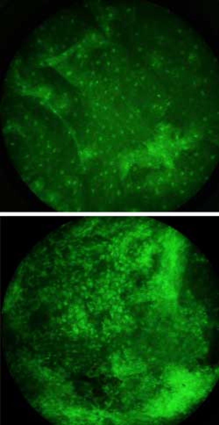
The nuclei of precancerous and cancerous cells are visibly distorted, so much so that the abnormal cells were visible even on the camera's small LCD screen. The team used tissue samples from resected tumors and cancer cells grown in a lab and compared them to the healthy tissue samples from patients' mouths.
"The dyes and visual techniques that we used are the same sort that pathologists have used for many years to distinguish healthy cells from cancerous cells in biopsied tissue," says study co-author Mark Pierce, a Rice faculty fellow in bioengineering. "But the tip of the imaging cable is small and rests lightly against the inside of the cheek, so the procedure is considerably less painful than a biopsy and the results are available in seconds instead of days."
Richards-Kortum, Rice's Stanley C. Moore Professor of Bioengineering as well as professor of electrical and computer engineering, heads a lab that has developed fluorescent dyes and targeted nanoparticles that let doctors zero in on the molecular hallmarks of cancer.
She says software could be written that would allow medical professionals who are not pathologists to use the device to distinguish healthy from non-healthy cells. The device could then be used for routine cancer screening and to help oncologists track how well patients were responding to treatment.
While a Rice spokesman says it could still be a while before additional testing and approvals come through to bring this technology to market, he adds that Richards-Kortum has already used it on herself, in front of an audience, showing the healthy nuclei of the cells in her mouth.