New technique uses virtual slides to view tissue in 3D
Researchers at the University of Leeds say their digital scanning system produces high-res images that can be rotated.


Today, pathologists and researchers must cut super-thin slices of tissue samples to view them on a microscope -- a labor-intensive process that renders 3D images created from hundreds of 2D sections prohibitively expensive.
Not to mention tedious to construct. Imagine if a single scene in Halo was presented as a series of 2D images one must perfectly align before getting the lay of, say, a single battleground.
Now, computer scientists and medical researchers at the University of Leeds in the United Kingdom say they've devised a novel workaround in the form of a digital scanning system that produces 3D views of tissue samples with almost no extra labor.
The team reports in the May issue of the American Journal of Pathology that its automated system turns batches of 2D slides into high-res digital images -- so-called virtual slides -- that are then easily aligned via image registration software. Researchers can then rotate and zoom in on these images.
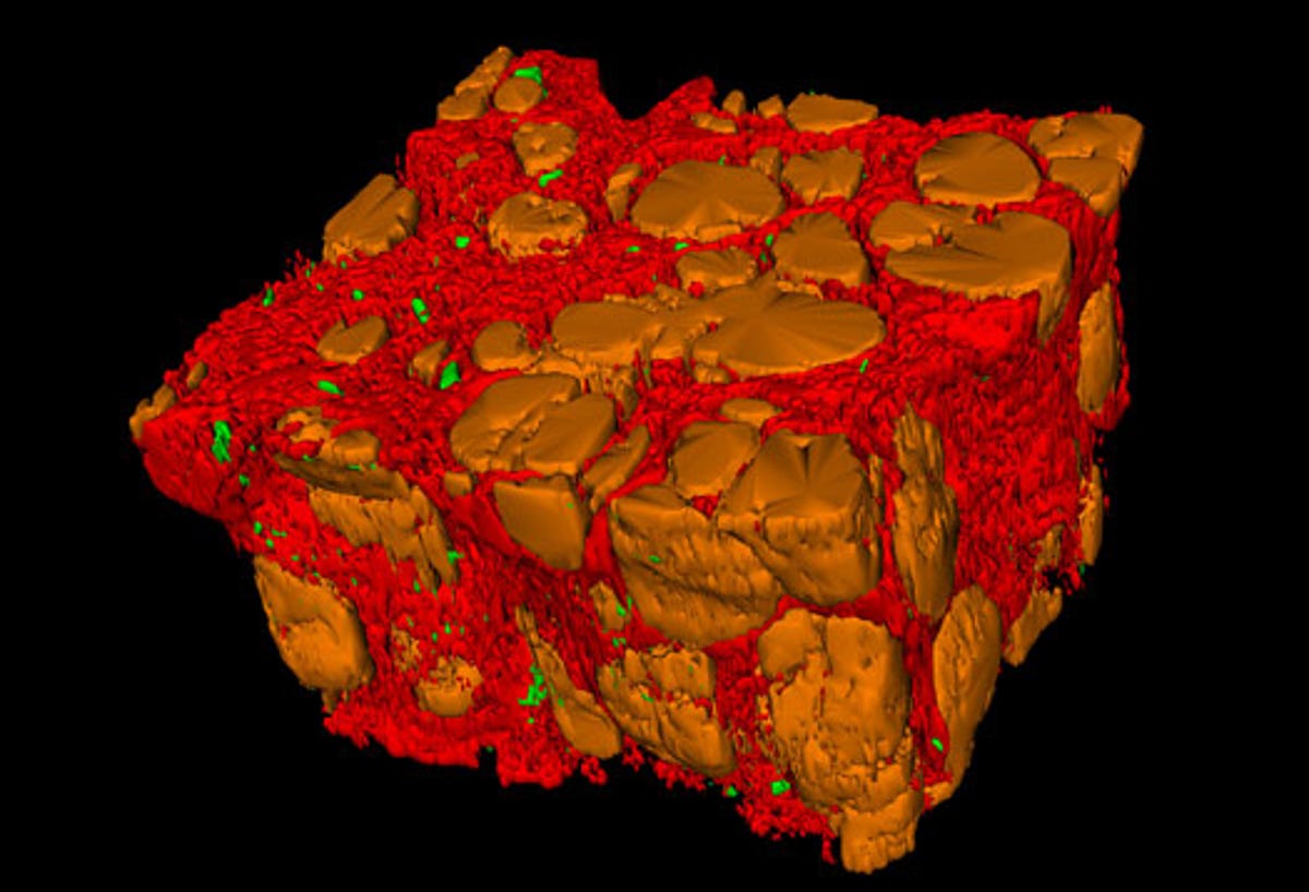
Being able to view tissue this way should help pathologists better view the shapes of the tissue they are studying, and thus glean more information about, say, the structures of developing organs and the branching of blood vessels to malignant tumors.
"Having a 3D view can often make a real difference," Derek Magee, who developed the software behind the system, said in a university news release. "For instance, if you want to understand how a system of blood vessels supplying a tumor connects up, you really need to see that in 3D, not as a series of separate 2D sections."
Magee and his colleagues have tested their approach on eight types of tissue using more than 13,000 virtual slides to get better views of liver disease, cancer, and embryos.
"Our virtual system means that users can look at the shape and structure of cells and the 'micro-architecture' of blood vessels and tumors on large tissue samples," said lead investigator Darren Treanor, a pathologist at the University of Leeds. "This can all be done without input from computing specialists."
What such a system will cost, and whether it will ever be practical in a clinical setting, remains to be seen. But it seems fair to posit that a system that automates the 3D rendering of 2D samples, at least when it comes to studying disease, will soon be worth the cost.