A 'Google Earth' approach to researching cells
Researchers stitch together more than 26,000 electron microscopy images to create a view of a zebrafish embryo that is 281 gigapixels with a resolution of 16 million pixels per inch.


Imagine being able to navigate our own biological tissue much in the way Google Earth allows us to zoom in on our own backyards. Only instead of mailboxes and fences, you could spot, say, rogue cancer cells.
Researchers out of Leiden University Medical Center in the Netherlands may be making such a future possible by stitching together molecular- and cellular-level images of biological tissues into truly gigantic composite images.
One such landscape -- of a zebrafish embryo -- consists of 26,000 images, is 281 gigapixels, and boasts a resolution of 16 million pixels per inch.
The researchers explain their advances to the imaging technique known as virtual nanoscopy in the Journal of Cell Biology. They were able to apply the technique to human dendritic cells, mouse embryonic cells, and the zebrafish embryo.
The Journal of Cell Biology, it turns out, had to revamp its own browser-based image-hosting platform, JCB DataViewer, just to provide access to the zebrafish embryo image. "If you can image it, you should be able to publish it," Liz Williams, the journal's executive editor, said yesterday in a statement.
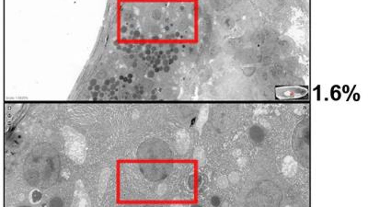
Now anyone can play around with the zebrafish embryo image using the platform, starting panned out to view the entire 1.5mm-long embryo and zooming in all the way to its molecular structures.
A key obstacle in imaging both molecular and cellular events simultaneously is working with the very different lengths, the researchers write: "Our approach employs standard transmission electron microscopy, rapid automated data collection, and stitching to create large virtual slides. It greatly facilitates correlative light-electron microscopy studies to relate structure and function and provides a genuine representation of ultrastructural events. The method is scalable as illustrated by slides up to 281 gigapixels in size."
Ultra indeed. How such a technique will ultimately be used remains to be seen, but subcellular inspection just got a bit grander.